-
 Melanocytoma of the disc
Dec 27 2023 by NIDHI PANWAR, MD FRCS Glasgow FNB FICO
Fundus photograph of an otherwise healthy 41 year old female , with recently detected diabetes mellitus and came for fundoscopy was Detected to have left eye optic disc melanocytoma .
Photographer: Nidhi Panwar, NMC Royal hospital, Sharjah , UAE
Condition/keywords: melanocytoma, optic disc
-
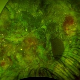 Advanced coats disease
Dec 27 2023 by NIDHI PANWAR, MD FRCS Glasgow FNB FICO
Fundus photograph of 6 year old otherwise healthy boy presented with right eye esotropia and poor vision with fundus picture depicting advanced exudative retinal disease suggestive of coats disease
Photographer: Nidhi Panwar, NMC Royal hospital, Sharjah , UAE
Condition/keywords: Coats disease, subretinal exudates
-
 Neovascularisation of disc
Dec 27 2023 by NIDHI PANWAR, MD FRCS Glasgow FNB FICO
Fundus photograph of 46 year old male, who is has poorly controlled Diabetes mellitus and has advanced proliferative diabetic retinopathy with Neovascularisation of disc right eye
Photographer: Nidhi Panwar, NMC Royal hospital, Sharjah , UAE
Condition/keywords: neovascularization of the disc (NVD), proliferative diabetic retinopathy (PDR)
-
 Morning Glory Disc Anomaly
Feb 12 2024 by NIDHI PANWAR, MD FRCS Glasgow FNB FICO
Fundus photograph of 43 year old male, hypertensive on medication, came for routine check up, and has been diagnosed to have poor vision left eye since childhood, denies any history of trauma. Vision left eye 6/18, Anterior segment normal, Fundus left eye shows excavated ,funnel-shaped optic nerve head, with central tuft of glial tissue obscuring the cup . The retinal vessels were seen emanating from the edge of disc in radial manner. In addition, the sectoral nasal retina shows localized area of hyperpigmented bony spicules like lesions. However, no history of nyctalopia or any other neurological disorder could be obtained.
Photographer: Nidhi Panwar, NMC Royal hospital, Sharjah , UAE
Imaging device: OPTOMAP
Condition/keywords: Morning Glory Anomaly, optic disc excavation
-
 Subhyaloid Hemorrhage
Oct 4 2025 by NIDHI PANWAR, MD FRCS Glasgow FNB FICO
33 Year old asymptomatic male with history of sudden blurring of right eye vision since 7 days ,
Photographer: Ms Ola
Imaging device: OPTOS
Condition/keywords: SUBHYALOID HEMORRHAGE
-
 Subhyaloid Hemorrhage
Oct 4 2025 by NIDHI PANWAR, MD FRCS Glasgow FNB FICO
33 Year old asymptomatic male with history of sudden blurring of right eye vision, after 5 days of observation by patient, vision getting more worse.
Photographer: Ms Ola
Imaging device: OPTOS
Condition/keywords: subhyaloid hemorrhage
-
 Subhyaloid Hemorrhage
Oct 4 2025 by NIDHI PANWAR, MD FRCS Glasgow FNB FICO
Pitting of ILM seen using green laser ( just before Yag hyaloidotomy)
Photographer: Ms Ola
Imaging device: OPTOS
Condition/keywords: Subhyaloid Hemorrhage
-
 Subhyaloid Hemorrhage
Oct 4 2025 by NIDHI PANWAR, MD FRCS Glasgow FNB FICO
Immediately post yag hyaloidotomy
Photographer: Ms Ola
Imaging device: OPTOS
Condition/keywords: subhyaloid hemorrhage
-
 Subhyaloid Hemorrhage
Oct 4 2025 by NIDHI PANWAR, MD FRCS Glasgow FNB FICO
3 days post yag hyaloidotomy ( clotted blood can be seen just below hyaloidotomy site )
Photographer: Ms Ola
Imaging device: OPTOS
Condition/keywords: subhyaloid hemorrhage
-
 Subhyaloid Hemorrhage
Oct 4 2025 by NIDHI PANWAR, MD FRCS Glasgow FNB FICO
Fully resolved subhyaloid hemorrhage post yag hyaloidotomy, 4 week later
Photographer: Ms Ola
Imaging device: optos
Condition/keywords: subhyaloid hemorrhage
-
 Stargardt Disease Autofluorescence
Oct 14 2025 by NIDHI PANWAR, MD FRCS Glasgow FNB FICO
Classical autofluorescence in Stargardt disease with foveal atrophy.
Photographer: Ms Amrutha Shaji Optometrist
Imaging device: Optos
Condition/keywords: Stargardt disease
-
 Emulsification of Silicon Oil Post Vitrectomy for Endophthalmitis
Oct 14 2025 by NIDHI PANWAR, MD FRCS Glasgow FNB FICO
Fundus picture post vitrectomy with silicon oil done for endophthalmitis shows early onset emulsification of silicon oil.
Photographer: Ms Safeena Salam Optometrist
Imaging device: OPTOS
Condition/keywords: endophthalmitis, silicon oil emulsification in vitreous cavity
-
 Progressive Increasing Emulsification of Silicon Oil Post Vitrectomy for Endophthalmitis
Oct 14 2025 by NIDHI PANWAR, MD FRCS Glasgow FNB FICO
Fundus picture post vitrectomy with silicon oil done for endophthalmitis shows early onset and progressive increasing emulsification of silicon oil.
Photographer: Ms Safeena Salam Optometrist
Imaging device: optos
Condition/keywords: endophthalmitis
-
 Choroidal Detachment Secondary to Pan Retinal Photocoagulation Left Eye
Oct 14 2025 by NIDHI PANWAR, MD FRCS Glasgow FNB FICO
Serous choroidal detachment noted temporally after two sittings of pan retinal photocoagulation in proliferative diabetic retinopathy left eye, which resolved in 1 week of observation and topical steroid drops.
Photographer: NIDHI PANWAR, NMC ROYAL HOSPITAL, SHARJAH
Imaging device: OPTOS
Condition/keywords: choroidal detachment, laser photocoagulation, pan retinal photocoagulation, proliferative diabetic retinopathy (PDR)
-
 Choroidal Detachment Secondary to Pan Retinal Photocoagulation Right Eye
Oct 14 2025 by NIDHI PANWAR, MD FRCS Glasgow FNB FICO
Serous choroidal detachment noted temporally after two sittings of pan retinal photocoagulation in proliferative diabetic retinopathy right eye, which resolved in 1 week of observation and topical steroid drops.
Photographer: NIDHI PANWAR, NMC ROYAL HOSPITAL, SHARJAH
Imaging device: optos
Condition/keywords: choroidal detachment, laser photocoagulation, pan retinal photocoagulation, proliferative diabetic retinopathy (PDR)
-
 Retinal Break With Cuff of Subretinal Fluid (Subclinical Retinal Detachment)
Nov 26 2025 by NIDHI PANWAR, MD FRCS Glasgow FNB FICO
Asymptomatic retinal break with subretinal fluid more than 2 DD in supero temporal quadrant
Photographer: NIDHI PANWAR
Imaging device: OPTOS
Condition/keywords: asymptomatic retinal break, asymptomatic retinal detachment, subretinal fluid
-
 Retinal Break With Cuff of Subretinal Fluid (Subclinical Retinal Detachment), Immediately Post Laser Barrage
Nov 26 2025 by NIDHI PANWAR, MD FRCS Glasgow FNB FICO
Immediately post laser barrage, case of asymptomatic retinal break with subretinal fluid more than 2 DD in supero temporal quadrant.
Photographer: NIDHI PANWAR
Imaging device: optos
Condition/keywords: asymptomatic retinal break, asymptomatic retinal detachment, BARRAGE LASER, subretinal fluid
-
 Dropped Iol With Glaucoma, Sclerosed Retinal Vessels and Retinal Hemorrhages
Jan 15 2026 by NIDHI PANWAR, MD FRCS Glasgow FNB FICO
82 year old female came to eye clinic with left eye pain and blurring of vision noted since past few months. She was found to have , vision of hand movements, high IOP with advanced glaucoma, sclerosed retinal vessels with retinal hemorrhage, and dislocated single piece lens in vitreous cavity.
Photographer: Dr Nidhi Panwar, NMC Royal hospital sharjah
Imaging device: optos
Condition/keywords: advanced stage glaucoma, dropped IOL, retinal hemorrhage, sclerosed vessels
A project from the American Society of Retina Specialists