-
 Chorioretinal Inflammation
Nov 1 2019 by Stephanie Burke
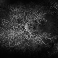
65-year-old woman with disseminated chorioretinal inflammation. Early frame of FA.
Photographer: Stephanie Burke, CRA, OCT-C
Imaging device: Optos
Condition/keywords: chorioretinal inflammations
-
 Chorioretinal Inflammation
Nov 1 2019 by Stephanie Burke
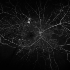
65-year-old woman with disseminated chorioretinal inflammation. Late frame of FA.
Photographer: Stephanie
Imaging device: Optos
Condition/keywords: chorioretinal inflammations
-
 Hamartoma
Nov 8 2019 by Stephanie Burke
Fundus Auto-fluorescence image of an 76-year-old man with hamartoma.
Photographer: Stephanie Burke, CRA, OCT-C
Imaging device: Optos
Condition/keywords: hamartoma
-
 Hamartoma
Nov 8 2019 by Stephanie Burke
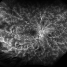
Early frame FA image of a 76-year-old man with hamartoma.
Photographer: Stephanie Burke, CRA, OCT-C
Imaging device: Optos
Condition/keywords: hamartoma
-
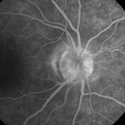 Papillophlebitis
Dec 23 2019 by Stephanie Burke
Fluorescein angiogram, mid-phase image of a 46-year-old woman with papillophlebitis.
Photographer: Stephanie Burke, CRA, OCT-C
Imaging device: Zeiss 450 Plus
Condition/keywords: optic disc edema, papillophlebitis
-
 Coats' Disease
Mar 2 2020 by Stephanie Burke
15-year-old female with Coats' disease.
Photographer: Stephanie Burke, CRA, OCT-C
Condition/keywords: Coats' disease, FA early phase, ultra-wide field imaging
-
 Uveal Effusion
Mar 25 2020 by Stephanie Burke
29-year-old male with uveal effusion. Patient was of short-stature with nanophthalmos.
Photographer: Stephanie Burke, CRA, OCT-C
Condition/keywords: chorioretinopathy, nanophthalmos, ultra-wide field imaging, uveal effusion, uveitis
-
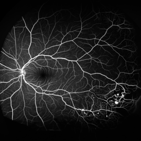
 Proliferative Diabetic Retinopathy
Apr 29 2020 by Stephanie Burke
Ultra-wide FA image of a 58-year-old man with DM.
Photographer: Stephanie Burke, CRA, OCT-C
Condition/keywords: ischemia, leakage, microaneurysms, neovascularization (NV), proliferative diabetic retinopathy (PDR), ultra-wide field imaging
-
 Central Retinal Vein Occlusion (CRVO)
Jun 19 2020 by Stephanie Burke
85-year-old male with ischemic CRVO.
Photographer: Stephanie Burke, CRA, OCT-C
Condition/keywords: central retinal vein occlusion (CRVO), ischemia, macular edema, ultra-wide field imaging
-
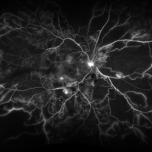
 PDR with Ischemia
Jul 7 2020 by Stephanie Burke
Early frame of a 45-year-old male with Type II diabetes.
Photographer: Stephanie Burke, CRA, OCT-C
Condition/keywords: FA early phase, ischemia, microaneurysms, neovascularization (NV), proliferative diabetic retinopathy (PDR), ultra-wide field imaging, venous beading
-
 Central Retinal Vein Occlusion
Feb 20 2021 by Stephanie Burke
Ultra wide-field FA image of a 22-year-old male with CRVO.
Photographer: Stephanie Burke, CRA ,OCT-C
Condition/keywords: central retinal vein occlusion (CRVO)
-
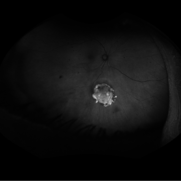
 Melanocytoma
Nov 3 2022 by Stephanie Burke
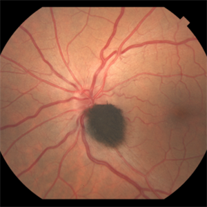
Topcon fundus photograph of a 49-year old female with a melanocytoma of the left eye.
Photographer: Stephanie Burke, CRA, OCT-C
Condition/keywords: melanocytoma
A project from the American Society of Retina Specialists