-
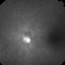
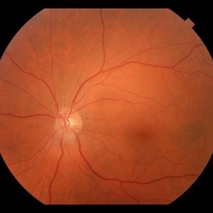 FP - Astrocytic Hamartoma
Oct 27 2019 by John S. King, MD
66-year-old white male without history of tuberous sclerosis was found to have an incidental, asymptomatic, translucent, retinal lesion with a few small telangiectatic vessels within it. The FA showed early hyperFL of these small vessels with prominent late leakage/staining. The OCT showed a retinal mass with a "moth eaten" appearance. Vision was 20/20 and the rest of the exam was unremarkable.
Photographer: Maisee Yang
Condition/keywords: astrocytic hamartoma
-
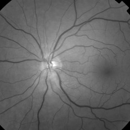 Red Free - Astrocytic Hamartoma
Oct 27 2019 by John S. King, MD
66-year-old white male without history of tuberous sclerosis was found to have an incidental, asymptomatic, translucent, retinal lesion with a few small telangiectatic vessels within it. The FA showed early hyperFL of these small vessels with prominent late leakage/staining. The OCT showed a retinal mass with a "moth eaten" appearance. Vision was 20/20 and the rest of the exam was unremarkable.
Photographer: Maisee Yang
Condition/keywords: astrocytic hamartoma
-
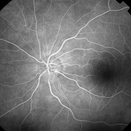 28 Sec (Laminar Flow) FA - Astrocytic Hemartoma
Oct 27 2019 by John S. King, MD
66-year-old white male without history of tuberous sclerosis was found to have an incidental, asymptomatic, translucent, retinal lesion with a few small telangiectatic vessels within it. The FA showed early hyperFL of these small vessels with prominent late leakage/staining. The OCT showed a retinal mass with a "moth eaten" appearance. Vision was 20/20 and the rest of the exam was unremarkable.
Photographer: Maisee Yang
Condition/keywords: astrocytic hamartoma
-
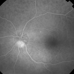 49 Sec FA - Astrocytic Hemartoma
Oct 27 2019 by John S. King, MD
66-year-old white male without history of tuberous sclerosis was found to have an incidental, asymptomatic, traslucent, retinal lesion with a few small telangiectatic vessels within it. The FA showed early hyperFL of these small vessels with prominent late leakage/staining. The OCT showed a retinal mass with a "moth eaten" appearance. Vision was 20/20 and the rest of the exam was unremarkable.
Photographer: Maisee Yang
Condition/keywords: astrocytic hamartoma
-
 2:30 FA - Astrocytic Hemartoma
Oct 27 2019 by John S. King, MD
66-year-old white male without history of tuberous sclerosis was found to have an incidental, asymptomatic, translucent, retinal lesion with a few small telangiectatic vessels within it. The FA showed early hyperFL of these small vessels with prominent late leakage/staining. The OCT showed a retinal mass with a "moth eaten" appearance. Vision was 20/20 and the rest of the exam was unremarkable.
Photographer: Maisee Yang
Condition/keywords: astrocytic hamartoma
-
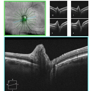 Juxtapapillay Astrocytic Hemartoma with Moth Eaten Appearance on OCT
Oct 27 2019 by John S. King, MD
66-year-old white male without history of tuberous sclerosis was found to have an incidental, asymptomatic, translucent, retinal lesion with a few small telangiectatic vessels within it. The FA showed early hyperFL of these small vessels with prominent late leakage/staining. The OCT showed a retinal mass with a "moth eaten" appearance. Vision was 20/20 and the rest of the exam was unremarkable.
Photographer: Maisee Yang
Condition/keywords: astrocytic hamartoma
A project from the American Society of Retina Specialists