-
 Paracentral Acute Middle Maculopathy
Oct 25 2019 by Gayathri Mohan
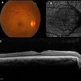
Multimodal images of follow up visit of a case of Paracentral acute middle maculopathy 2 months post oral steroid therapy. Color fundus photograph(A) showing resolution of whitish retinal lesions . OCT and OCTA (B,C) also show resolution of lesions post treatment.
Photographer: Akshar Soni
Imaging device: Heidelberg, Nidek
Condition/keywords: fundus albipunctatus, optical coherence tomography (OCT), paracentral acute middle maculopathy
-
 Paracentral Acute Middle Maculopathy
Oct 25 2019 by Gayathri Mohan
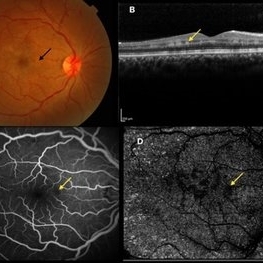
Multimodal images of a case of a 29-year-old female with paracentral acute middle maculopathy. A-color fundus photograph showing multiple confluent white retinal patches. B- On OCT the acute lesions of PAMM characteristically appear as placoid, hyperreflective bands at the level of the INL C-Fundus fluorescein angiography showing a capillary nonperfusion area D-flow void areas in deep capillary plexus
Photographer: Akshar Soni
Imaging device: Heidelberg, Nidek
Condition/keywords: fundus albipunctatus, optical coherence tomography (OCT), paracentral acute middle maculopathy
A project from the American Society of Retina Specialists