-
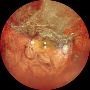
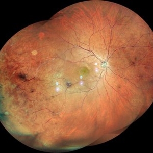
 LE WIDEFIELD FUNDUS PHOTOGRAPH OF HYPERTENSIVE RETINOPATHY GRADE IV
Sep 27 2023 by ANKIT JAIN
WIDEFIELD IMAGE OF RE OF 14 YEARS OLD MALE RECENTLY DIAGNOSED WITH MALIGNANT HYPERTENSION SHOWING MACULAR STAR, DISC EDEMA IN A CASE OF HYPERTENSIVE RETINOPATHY GRADE IV.
Photographer: DR ANKIT JAIN
Imaging device: MIRANTE
Condition/keywords: Disc Edema, hypertensive retinopathy, macular star
-
 RE WIDEFIELD FUNDUS PHOTOGRAPH OF HYPERTENSIVE RETINOPATHY GRADE IV
Sep 27 2023 by ANKIT JAIN
WIDEFIELD IMAGE OF RE OF 14 YEARS OLD MALE RECENTLY DIAGNOSED WITH MALIGNANT HYPERTENSION SHOWING MACULAR STAR, DISC EDEMA IN A CASE OF HYPERTENSIVE RETINOPATHY GRADE IV.
Photographer: DR ANKIT JAIN
Imaging device: MIRANTE
Condition/keywords: Disc Edema, hypertensive retinopathy, macular star
-
 Malignant Hypertension
Sep 28 2023 by ANKIT JAIN
Widefield photograph of LE of a 13 year old male with malignant hypertension
Photographer: Dr. Ankit Jain
Imaging device: Mirante
Condition/keywords: hypertensive retinopathy, malignant hypertension
-
 Malignant Hypertension
Sep 28 2023 by ANKIT JAIN
Fundus photograph of LE of a 13 year old male with malignant hypertension
Photographer: Dr. Ankit Jain
Imaging device: Mirante
Condition/keywords: hypertensive retinopathy, malignant hypertension
-
 Malignant Hypertension
Sep 28 2023 by ANKIT JAIN
Fundus photograph of RE of a 13 year old male with malignant hypertension
Photographer: Dr. Ankit Jain
Imaging device: Mirante
Condition/keywords: hypertensive retinopathy, malignant hypertension
-
 LARGE OPERCULATED HOLE
Oct 16 2023 by ANKIT JAIN
Left Eye Montage of 50 years old male with high myopia with large operculated hole
Photographer: DR ANKIT JAIN
Imaging device: MIRANTE
Condition/keywords: High Myopia, myopia, operculated retinal hole
-
 SUB HYALOD HEMORRHAGE
Oct 18 2023 by ANKIT JAIN
WIDEFIELD FUNDUS IMAGE OF RIGHT EYE OF 53 YEAR OLD MALE, KNOWN CASE OF DIABETES MELLITUS TYPE 2 FROM 3 YEARS WITH SUB-HYALOID HEMORRGAE IN CASE OF PROLIFERATIVE DIABETIC RETINOPATHY
Photographer: DR ANKIT JAIN
Imaging device: MIRANTE
Condition/keywords: diabetes, proliferative diabetic retinopathy (PDR)
-
 Macular BRVO
Oct 22 2023 by ANKIT JAIN
LE fundus photo of 62 year old male with macular branch retinal vein occlusion with macular edema
Photographer: DR ANKIT JAIN
Imaging device: MIRANTE
Condition/keywords: macular branch retinal vein occlusion (BRVO), macular edema
-
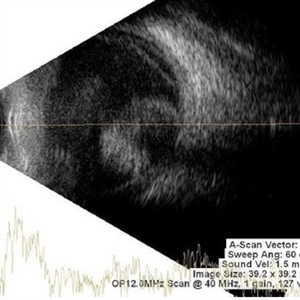
 POSTERIOR SCLERITIS
Nov 1 2023 by ANKIT JAIN
USG B SACN image showing typical T-sign in axial horizontal view with increased thickening of the sclero-choroidal complex suggestive of posterior scleritis
Photographer: DR ANKIT JAIN
Condition/keywords: B scan ultrasound, posterior scleritis, ULTRASOUND
-
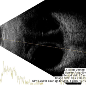
 Posterior Vitreous Detachment
Nov 1 2023 by ANKIT JAIN
USG B SCAN image showing membranous echoes with low to moderate spikes with free after movements with no attachment to disc suggestive of posterior vitreous detachment.
Photographer: DR ANKIT JAIN
Condition/keywords: B scan ultrasound, posterior vitreous detachment, PVD, ultrasound
-
 Choroidal Hemangioma
Nov 1 2023 by ANKIT JAIN
USG B SCAN image showing hyperechogenic mass like lesion with uniform height of spikes through out the mass likely suggestive of choroidal hemangioma
Photographer: DR ANKIT JAIN
Condition/keywords: B scan ultrasound, choroidal hemangioma, ultrasound
-
 Retinoblastoma
Nov 1 2023 by ANKIT JAIN
USG B SCAN image showing hyperechogenic mass lesion with moderate spikes with restricted after movements on dynamic scan. In between high spikes noted suggestive of calcification in a case of Retinoblastoma
Photographer: DR ANKIT JAIN
Condition/keywords: B scan ultrasound, retinoblastoma, ultrasound
-
 Choroidal Melanoma
Nov 1 2023 by ANKIT JAIN
USG B SCAN image showing mass echoes with internal homogeneity with attenuating spikes in a decrescendo pattern likely suggestive of choroidal melanoma.
Photographer: DR ANKIT JAIN
Condition/keywords: B scan ultrasound, CHoroidal melanoma, ultrasound
-
 Phthisis Bulbi
Nov 1 2023 by ANKIT JAIN
USG B SCAN image showing multiple dot echos with mild to moderate spikes with free after movements on dynamic scan suggestive of vitreous degeneration, decreased axial length notedand loss of the normal shape of globe suggestive of phthisis bulbi
Photographer: DR ANKIT JAIN
Condition/keywords: B scan ultrasound, phthisis bulbi
-
 Asteroid Hyalosis
Nov 1 2023 by ANKIT JAIN
B sacn ultrasound image showing hyperechoic echoes with mild to moderate spikes with free after movements on dynamic scan suggestive of asteroid hyalosis.
Photographer: DR ANKIT JAIN
Condition/keywords: asteroid hyalosis, B scan ultrasound
-
 Superior retinal detachment with break
Nov 20 2023 by ANKIT JAIN
Widefield fundus image of LE showing superior retinal detachment with break
Photographer: Dr Ankit Jain
Imaging device: MIRANTE
-
 CRAO
CRAO
Jan 8 2024 by ANKIT JAIN
RIGHT EYE FUNDUS IMAGE OF A 68 YEARS OLD MALE WITH SUDDEN LOSS OF VISION, WHO IS A KNOWN CASE OF HYPERTENSION FOR 15 YEARS
Photographer: Dr Ankit Jain
Condition/keywords: central retinal artery occlusion (CRAO), cherry red spot
-
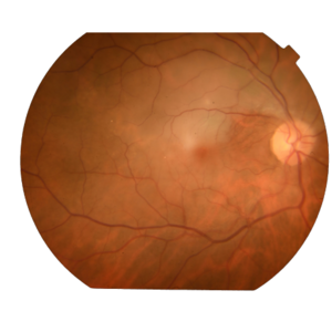
 OLD MACULAR BRVO
Jan 8 2024 by ANKIT JAIN
LE FUNDUS IMAGE OF 58 YEARS OLD MALE WITH OLD MACULAR BRVO SHOWING SCLEROSED VESSEL
Photographer: Dr Ankit Jain
Condition/keywords: macular branch retinal vein occlusion (BRVO)
-
 BRVO WITH SCLEROSED VESSELS: RE
Jan 8 2024 by ANKIT JAIN
RIGHT EYE FUNDUS IMAGE OF 58 YEARS OLD MALE WITH OLD BRVO HAVING SCLEROSED VESSELS
Photographer: Dr Ankit Jain
Imaging device: MIRANTE
Condition/keywords: branch retinal vein occlusion (BRVO)
-
 LASER PRP: LE
Jan 8 2024 by ANKIT JAIN
LEFT EYE WIDEFILED FUNDUS PHOTO OF 38 YEARS OLD FEMALE WITH TYPE 1 DIABETES MELLITUS HAVING WITH 360 DEGREE LASER PRP
Photographer: DR ANKIT JAIN
Imaging device: MIRANTE
Condition/keywords: Diabetes, pan-retinal photocoagulation (PRP), PRP
-
 VITREOUS HEMORRHAGE WITH 360 DEGREE LASER PRP: RE
Jan 8 2024 by ANKIT JAIN
RIGHT EYE WIDEFILED FUNDUS PHOTO OF 38 YEARS OLD FEMALE WITH TYPE 1 DIABETES MELLITUS HAVING VITREOUS HEMORRHAGE WITH 360 DEGREE LASER PRP
Photographer: Dr Ankit Jain
Imaging device: MIRANTE
Condition/keywords: Diabetes, pan-retinal photocoagulation (PRP), PRP, vitreous hemorrhage
-
 Choroidal Detachment
Jan 12 2024 by ANKIT JAIN
Membranous echoes with moderate to high spikes with restricted after movements suggestive of choroidal detachment.
Photographer: Dr Ankit Jain
Condition/keywords: choroidal detachment
-
 Choroidal Mass
Mar 4 2024 by ANKIT JAIN
Left eye color photo montage of 39 year old female with sub retinal mass in nasal quadrant with hemorrhages and subretinal fluid with inferior retinal detachment.
Photographer: Dr Ankit Jain
Imaging device: MIRANTE
Condition/keywords: choroidal mass
-
 Choroidal Mass
Mar 4 2024 by ANKIT JAIN
RE color photo montage of right eye of 48 year old with sub retinal hemorrhage with sub retinal fluid at level of fovea.
Photographer: Dr Ankit Jain
Imaging device: MIRANTE
Condition/keywords: macroaneurysm, retinal arterial macroaneurysm
A project from the American Society of Retina Specialists