-
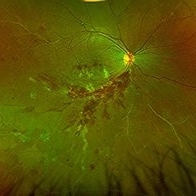
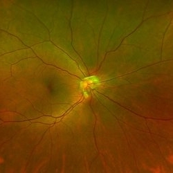
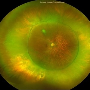
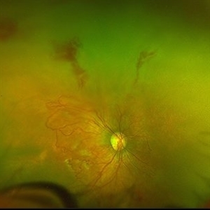
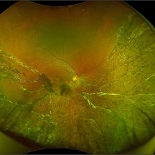 Self-Settled Retinal Detachment
Aug 21 2023 by rahul saradge
35 YEARS OLD MALE SELF SETTLED INF RD, CRA, SCLEROSED VESSELS
Photographer: Rekha Pathak, Isha Netralaya
Condition/keywords: SELF SETTLED RETINAL DETACHMENT
-
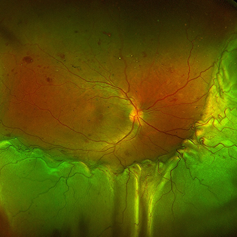
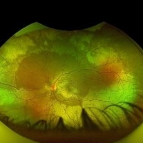 Berlin's Edema
Aug 21 2023 by rahul saradge
19 YEAR OLD MALE , HISTORY OF TRAUMA FEW HOURS BEFORE IMAGING
Photographer: Ramjan Shaikh, Isha Netralaya
Imaging device: Optos
Condition/keywords: Berlin's edema
-
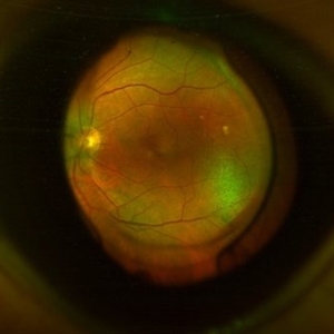
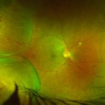
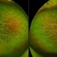
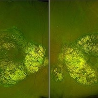 Central Areolar Choroidal Dystrophy
Aug 21 2023 by rahul saradge
54 year old female well circumscribed, bilateral and symmetrical lesion with loss of retinal and choroidal tissue in the macular area.
Photographer: Sushil Zende, Isha Netralaya
Imaging device: Optos
Condition/keywords: central areolar choroidal dystrophy (CACD)
-
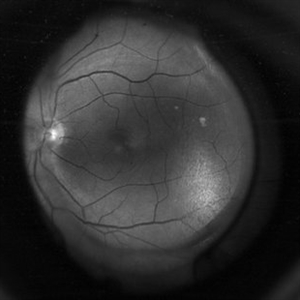
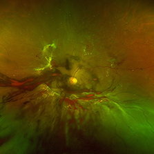
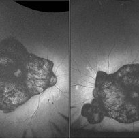 Central Areolar Choroidal Dystrophy
Aug 21 2023 by rahul saradge
54 year old female well circumscribed, bilateral and symmetrical lesion with loss of retinal and choroidal tissue in the macular area.
Photographer: Sushil Zende, Isha Netralaya
Imaging device: Optos
Condition/keywords: autofluorescence imaging, central areolar choroidal dystrophy (CACD)
-
 BRVO and Perivascular Sheathing
Aug 21 2023 by rahul saradge
30 years old male with BRVO and perivascular sheathing .
Photographer: Dhruvakshi Kharade, Isha Netralaya
Imaging device: Optos
Condition/keywords: BRVO, Perivascular Sheathing
-
 CMV Retinitis with Shallow RD
Aug 21 2023 by rahul saradge
47 YEAR OLD MALE HAVING CMV RETINITIS WITH SHALLOW RD, VITRITIS
Photographer: Hitesh Rawlani , Isha Netralaya
Condition/keywords: cmv retinits with shallow RD
-
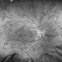
 Proliferative diabetic retinopathy
Aug 22 2023 by rahul saradge
39 year old with unstable PDR s/p PRP , vitreous hemorrhage.
Photographer: Aniket Pednekar , Isha Netralaya
Condition/keywords: PRP, red-free, vitreous blood
-
 CRVO, Disc Edema and Macular Thickening
Aug 22 2023 by rahul saradge
68 year old male with CRVO, disc edema and macular thickening (red free)
Photographer: Neha Singh ,Isha Netralaya
Condition/keywords: central retinal vein occlusion (CRVO), disc edema, red-free
-
 Bergmeister's Papilla
Sep 1 2023 by rahul saradge
Bergmeister's papilla
Photographer: Ajay Butta ,Isha Netralaya
Condition/keywords: Bergmeister's Papillae, Optos
-
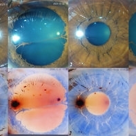 Persistent Pupillary Membrane
Oct 4 2023 by rahul saradge
45 year old female present with a complaint of recurrently something moving in front of the right eye. slit lamp examination suggestive of PPM with prominent collarette.
Photographer: Meenakshi Sonawane (Intern), Isha Netralaya
Imaging device: Appaswamy Dynamic imaging system
Condition/keywords: persistent pupillary membrane
-
 Antero-Posterior Glance
Nov 5 2023 by rahul saradge
image through the principle axis with visibility of all structure in pathway .
Photographer: Optom Rahul , Isha Netralaya
Condition/keywords: optical, Optos, posterior chamber intraocular lens (PCIOL)
-
 Antero-Posterior Glance
Nov 5 2023 by rahul saradge
image through the principle axis with visibility of all structure in pathway .
Photographer: Optom Rahul , Isha Netralaya
Condition/keywords: IOL, optos, retina
-
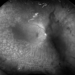
 Scleral buckle indent s/p retina surgery
Dec 13 2023 by rahul saradge
Scleral buckle indent s/p retina surgery
Photographer: Saloni Mishra , Isha Netralaya.
Imaging device: optos
Condition/keywords: Optos, Retina buckle, retina surgery, scleral buckle, ultra-wide field imaging
-
 Combined Pathology
Oct 26 2024 by rahul saradge
53 year old male patient was presented with a complaints of diminished vision in LE since 1 month. The BCVA in RE was 6/36p and LE was CF 1/2m. Ocular dilated examination showed RE temporal CD with ?CRVO,OIS and OS showed TRD and old Hemi CRVO. Patient was injected with PST tricot followed by PRP laser at an interval of 1 week. Patient improved to BCVA 6/9.
Photographer: Aishwarya Bangar Isha Netralaya Thane
Imaging device: optos
Condition/keywords: choroidal detachment, crvo, ois, optos, pan retinal photocoagulation, tractional retinal detachment
-
 Diabetic Retinopathy
Oct 26 2024 by rahul saradge
Patient was presented to clinic with a complaint of progressive DOV and retinal evaluation showed PDR.
Photographer: Lokesh Dukare ,Isha Netralaya Thane
Imaging device: optos
Condition/keywords: diabetic retinopathy, optos, PDR, retina, Retinal Detachment with Vitreous Hemorrhage
-
 Aggressive ROP
Oct 26 2024 by rahul saradge
Premature baby referred for ROP evaluation. HALF ZONE 1 was only vascularised, Patient was given Inj Anti-Vegf followed by ROP Laser after 1 week.
Photographer: Ankita Choudhary ,Isha Netralaya Thane
Imaging device: optos
Condition/keywords: aggresive retinopathy of prematurity, optos, retinopathy of prematurity
-
 Aggressive ROP
Oct 26 2024 by rahul saradge
Premature baby referred for ROP evaluation. HALF ZONE 1 was only vascularised, Patient was given Inj Anti-Vegf follwed by ROP Laser after 1 week
Photographer: Neha Choudhary,Isha Netralaya Thane
Imaging device: optos
Condition/keywords: aggressive posterior retinopathy of prematurity (APROP), retinopathy of prematurity
-
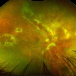
 Exudative Retinal Detachment
Oct 26 2024 by rahul saradge
41y/M, k/c/o TYPE 1 DM DOV SINCE 1 WEEK OU H/O TYPHOID 2 WEEKS BACK Here is the image showing Exudative RD in Right Eye We Planned PRP laser for this patient, advised him Carotid Doppler and 2D ECHO
Photographer: Anagha Wakode, Isha Netralaya Thane
Imaging device: optos
Condition/keywords: choroidals, exudative Retinal detachment
-
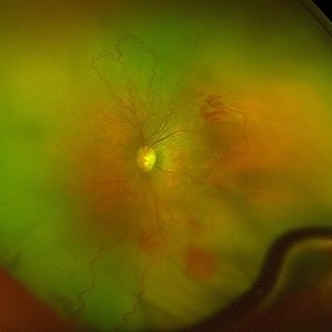
 Advanced RP
Nov 5 2024 by rahul saradge
A man, 58, arrived complaining of BOV for both near and distance vision in both eyes, with a 6/9 BCVA in each eye. For a year, the patient had been taking medication for both diabetes and hypertension. In both eyes, the dilated ophthalmoscopic retina revealed waxy disc pallor paired with bony spicules in the mid-periphery. The patient was prescribed spectacles and given counseling regarding the nature of the illness.
Photographer: Lokesh Dukare ,Isha Netralaya Thane
Imaging device: optos
Condition/keywords: bone spicule, optic disc pallor, Optos, Retinitis Pigmentosa
A project from the American Society of Retina Specialists