-
 Retinal Arterial Macroaneurysm
Apr 8 2023 by Yousef A Fouad, MD, FRCS (Glasg.)
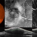
Multimodal imaging of a retinal arterial macroaneurysm in the right eye of a 73-year-old male patient with uncontrolled hypertension. Fundus photography shows hemorrhage surrounding an arterial branch of the upper temporal arcade. Optical coherence tomography (OCT) through the lesion shows inner retinal hyperreflectivity with back shadowing, and adjacent cystoid macular edema in the outer retina. En face OCT centered on the lesion delineates the fusiform dilatation of the affected vessel, and OCT angiography confirms the presence of blood flow within the aneurysmal dilatation.
Photographer: Yousef Fouad, Ain Shams University, Egypt
Condition/keywords: arteriolar macroaneurysm, enface imaging, macroaneurysm, macroarterial aneurysm, OCT Angiography, OCTA
A project from the American Society of Retina Specialists