-
 27-Gauge Vitrectomy
Aug 3 2013 by Yusuke Oshima, MD, PhD
A high-performance 27-gauge vitrectomy system (27+) was used for treating a case with primary rhegmatogenous retinal detachment complicated with macular hole.
Condition/keywords: macular hole, video, vitrectomy
-
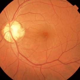
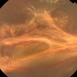
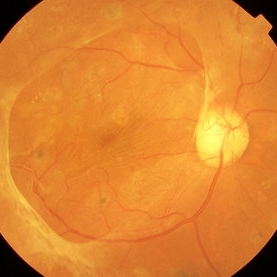
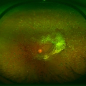
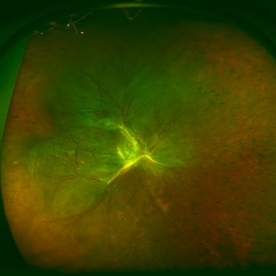 Traction Retinal Detachment Associated With Proliferative Diabetic Retinopathy
Aug 3 2013 by Yusuke Oshima, MD, PhD
Wide-angle fundus photograph of a 47-year-old man with massive fibrovascular proliferation and traction retinal detachment due to proliferative diabetic retinopathy.
Condition/keywords: tractional retinal detachment
-
 Retinal Capillary Hemangioma
Mar 21 2013 by Yusuke Oshima, MD, PhD
A peripheral retinal capillary hemangioma.
Photographer: Yusuke Takada, Osaka University Graduate School of Medicine
-
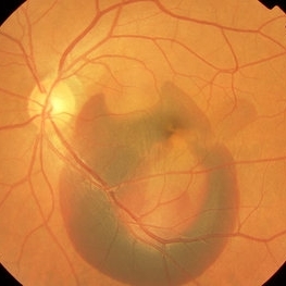
 Retinal Detachment Due to Traumatic Retinal Breaks
Mar 21 2013 by Yusuke Oshima, MD, PhD
Focal retinal detachment secondary to traumatic retinal breaks.
Photographer: Yusuke Takada, Osaka University Graduate School of Medicine
-
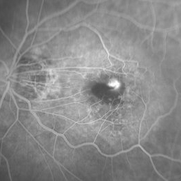
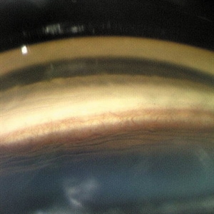
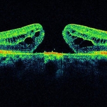 Idiopathic Macular Hole
Mar 21 2013 by Yusuke Oshima, MD, PhD
SD-OCT finding of a idiopathic macular hole.
Photographer: Yusuke Takada, Osaka University Graduate School of Medicine
Condition/keywords: macular hole
-
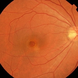 Idiopathic Macular Hole
Mar 21 2013 by Yusuke Oshima, MD, PhD
Fundus photograph of a idiopathic macular hole.
Photographer: Yusuke Takada, Osaka University Graduate School of Medicine
Condition/keywords: macular hole
-
 Epiretinal Membrane Proliferation
Mar 21 2013 by Yusuke Oshima, MD, PhD
Epiretinal membrane proliferation
Photographer: Yusuke Takada, Osaka University Graduate School of Medicine
Condition/keywords: epiretinal membrane (ERM)
-
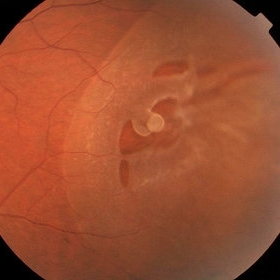
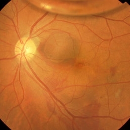
 Pit Macular Syndrome
Mar 21 2013 by Yusuke Oshima, MD, PhD
Fundus photograph of a 38-year-old man with macular detachment associated with an optic disc pit.
Photographer: Yusuke Takada, Osaka University Graduate School of Medicine
Condition/keywords: congenital optic nerve pit
-
 Extra-Macula Subretinal Hemorrhage
Mar 21 2013 by Yusuke Oshima, MD, PhD
Fundus photograph of an 86-year-old woman with an extra-macula subretinal hemorrhage associated with polypoidal choroidal vasculopathy.
Photographer: Yusuke Takada, Osaka University Graduate School of Medicine
-
 Submacular Hemorrhage After Pneumatic Displacement
Mar 21 2013 by Yusuke Oshima, MD, PhD
Fundus photograph demonstrates an effective displacement of the subretinal hemorrhage from the fovea.
Photographer: Yusuke Takada, Osaka University Graduate School of Medicine
Condition/keywords: submacular hemorrhage
-
 Submacular Hemorrhage Before Treatment
Mar 21 2013 by Yusuke Oshima, MD, PhD
Fundus photograph of an 83-year-old man with a submacular hemorrhage due to polypoidal choroidal vasculopathy.
Photographer: Yusuke Takada, Osaka University Graduate School of Medicine
Condition/keywords: submacular hemorrhage
-
 Myopic CNV
Mar 21 2013 by Yusuke Oshima, MD, PhD
Fluorescein angiogram illustrates a subfoveal myopic CNV.
Photographer: Yusuke Takada, Osaka University Graduate School of Medicine
Condition/keywords: myopic choroidal neovascularization (CNV)
-
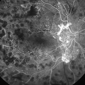
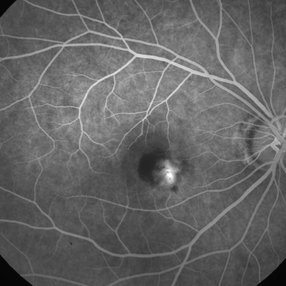 Idiopathic Choroidal Neovascularization
Mar 21 2013 by Yusuke Oshima, MD, PhD
Fluorescein angiogram of a 36-year-old woman with an idiopathic juxtafoveal CNV.
Photographer: Yusuke Takada, Osaka University Graduate School of Medicine
Condition/keywords: choroidal neovascularization (CNV)
-
---thumb.jpg/image-square;max$300,300.ImageHandler) Subconjunctival Air Bubbles
Subconjunctival Air Bubbles
Mar 21 2013 by Yusuke Oshima, MD, PhD
Slit lamp photograph demonstrates subconjunctival air bubbles, which is attributed to incomplete self-sealing of sclerotomies in a 25-gauge microincision vitrectomy surgery.
Photographer: Yusuke Takada, Osaka University Graduate School of Medicine
Condition/keywords: complication
-
 Falciform Retinal Detachment Associated with Toxocariasis (Photo-2)
Mar 21 2013 by Yusuke Oshima, MD, PhD
Fundus photograph of a 6-year-old boy with a falciform retinal detachment suspiciously associated with toxocariasis.
Photographer: Yusuke Takada, Osaka University Graduate School of Medicine
-
 Falciform Retinal Detachment Associated with Toxocariasis (Photo-1)
Mar 21 2013 by Yusuke Oshima, MD, PhD
Fundus photograph of a 6-year-old boy with a falciform retinal detachment suspiciously associated with toxocariasis.
Photographer: Yusuke Takada, Osaka University Graduate School of Medicine
-
 Regression of Iris Neovascularization After Intravitreal Bevacizumab
Mar 21 2013 by Yusuke Oshima, MD, PhD
Remarkable regression of iris neovascularization one day after intravitreal bevacizumab.
Photographer: Yusuke Takada, Osaka University Graduate School of Medicine
Condition/keywords: intravitreal bevacizumab, iris neovascularization
-
 Advanced Stage of Neovascular Glaucoma
Mar 21 2013 by Yusuke Oshima, MD, PhD
An 82-year-old man with a advanced stage of neovascular glaucoma. A slit-lamp photograph illustrates iris ectropion with prominent iris neovascularization.
Photographer: Yusuke Takada, Osaka University Graduate School of Medicine
Condition/keywords: neovascular glaucoma
-
 Angle Neovascularization
Mar 21 2013 by Yusuke Oshima, MD, PhD
Angle neovascularization due to ischemic CRVO.
Photographer: Yusuke Oshima, MD, PhD, Osaka University Graduate School of Medicine
Condition/keywords: angle neovascularization, gonioscopy
-
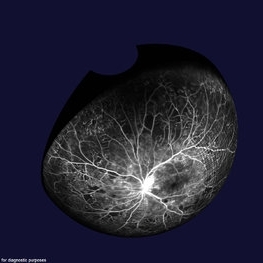
 Fluorescein Angiogram of the Case with Proliferative Diabetic Retinopathy
Mar 21 2013 by Yusuke Oshima, MD, PhD
Fluorescein angiography demonstrates a prominent neovascular network at the disc with an enlarged avascular zone at the macula.
Photographer: Yusuke Takada, Osaka University Graduate School of Medicine
-
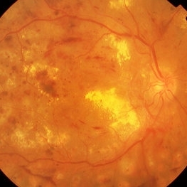 Proliferative Diabetic Retinopathy
Mar 21 2013 by Yusuke Oshima, MD, PhD
Fundus photograph of a 62-year-old man with PDR.
Photographer: Yusuke Takada, Osaka University Graduate School of Medicine
-
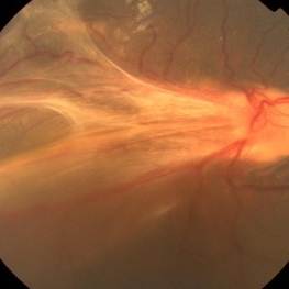
 Diabetic Traction Retinal Detachment
Mar 21 2013 by Yusuke Oshima, MD, PhD
Fundus photograph of a 42-year-old man with TRD due to PDR.
Photographer: Taku Wakabayashi, MD, Osaka University Graduate School of Medicine
-
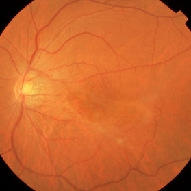
 Diabetic Macular Heterotopia
Mar 21 2013 by Yusuke Oshima, MD, PhD
Fundus photograph demonstrates a macular heterotopia secondary to the fibrovascular mambrane formation.
Photographer: Yusuke Takada, Osaka University Graduate School of Medicine
-
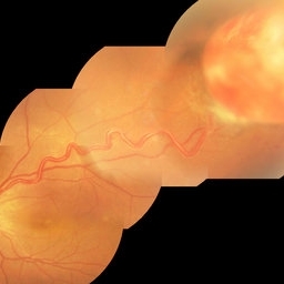
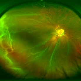
 3D-Image of Fluorescein Angiogram in a PDR Case
Mar 21 2013 by Yusuke Oshima, MD, PhD
A composite 3D-image of the fluorescein angiogram of the same PDR case.
Photographer: Yusuke Takada, Osaka University Graduate School of Medicine
Imaging device: OPTOS 200Tx
-
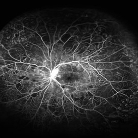 Fluorescein Angiogram of Proliferative Diabetic Retinopathy
Mar 21 2013 by Yusuke Oshima, MD, PhD
Wide-field fluorescein angiogram demonstrating prominent leakage from the NVD and scattered non-perfusion areas at the mid-pheriphery.
Photographer: Yusuke Takada, Osaka University Graduate School of Medicine
-
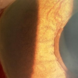
---thumb.jpg/image-square;max$300,300.ImageHandler) Rhegmatogeous Retinal Detachment
Rhegmatogeous Retinal Detachment
Mar 21 2013 by Yusuke Oshima, MD, PhD
Wide-field fundus photograph of a 58-year-old woman with a macula-involved bullous retinal detachment due to a superotemporal retinal break.
Photographer: Yusuke Takada, Osaka University Graduate School of Medicine
Imaging device: OPTOS 200Tx
Condition/keywords: bullous retinal detachment
-
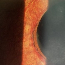
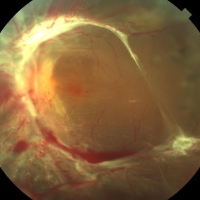
 Rhegmatogeous Retinal Detachment
Mar 21 2013 by Yusuke Oshima, MD, PhD
Wide-field fundus photograph of a 38-year-old woman with a macula-involved retinal detachment due to a tiny break localized around the edge of a lattice degeneration.
Photographer: Yusuke Takada, Osaka University Graduate School of Medicine
Imaging device: OPTOS 200Tx
-
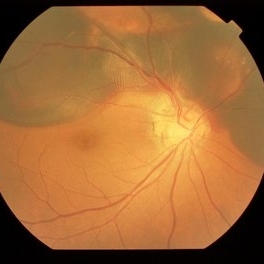
 Diabetic Traction Retinal Detachment
Mar 21 2013 by Yusuke Oshima, MD, PhD
Wide-field fundus photograph of a 56-year-old man with tractional retinal detachment due to proliferative diabetic retinopathy.
Photographer: Yusuke Takada, Osaka University Graduate School of Medicine
Imaging device: OPTOS 200Tx
A project from the American Society of Retina Specialists