-
 Asteroid Hyalosis
Feb 15 2023 by Anand Temkar
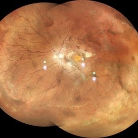
Fundus photo of LE of a 67 year old male with asteroid hyalosis.
Photographer: Dr.Anand Temkar
Imaging device: Mirante
Condition/keywords: asteroid hyalosis
-
 Anaemic Retinopathy
Sep 13 2023 by Anand Temkar
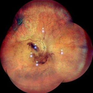
Wide field image of the RE of a 35 year old male patient showing Roth's spots in all four quadrants and venous tortuosity in a case of Anaemic Retinopathy.
Photographer: Dr.Anand Temkar- Retina Foundation, Ahmedabad
Imaging device: Mirante
Condition/keywords: anaemic retinopathy, roth spots
-
 Vitreous Haemorrhage with pre-retinal haemorrhage in case of a proliferative diabetic retinopathy
Sep 14 2023 by Anand Temkar
Wide field image of the right eye of a 53 year old male patient showing vitreous haemorrhage with pre-retinal haemorrhage in case of a proliferative diabetic retinopathy.
Photographer: Dr.Anand Temkar - Retina foundation, Ahmedabad
Imaging device: Mirante
Condition/keywords: pre-retinal hemorrhage, vitreous hemorrhage
-
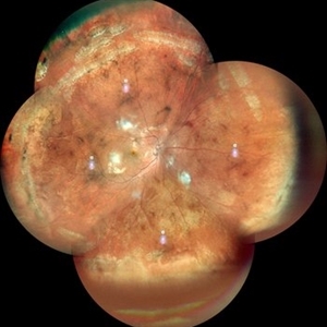 Pathological Myopia
Oct 18 2023 by Anand Temkar
RE widefield color photo montage of a 25 years old male showing lattice degenerations in periphery in a case of pathological myopia.
Photographer: Dr.Anand Temkar- Retina Foundation, Ahmedabad
Imaging device: Mirante
Condition/keywords: high myopia, lattice degeneration
-
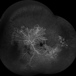 Old BRVO
Oct 18 2023 by Anand Temkar
LE widefield FA montage of a 68 years old male with history of Old BRVO, showing peripheral capillary non perfusion and some temporal laser marks (staining ).
Photographer: Dr.Anand Temkar- Retina Foundation, Ahmedabad
Imaging device: Mirante
Condition/keywords: branch retinal vein occlusion (BRVO), capillary nonperfusion
-
 Pathological Myopia
Oct 18 2023 by Anand Temkar
RE widefield CF montage of a 24 year old male with pathological myopia showing multiple lattice degenerations in periphery along with holes.
Photographer: Dr.Anand Temkar- Retina Foundation, Ahmedabad
Imaging device: Mirante
Condition/keywords: high myopia, holes, lattice degeneration
-
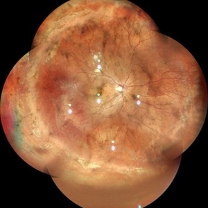
 Pathological Myopia
Oct 18 2023 by Anand Temkar
LE widefield CF montage of a 24 year old male with pathological myopia showing multiple lattice degenerations in periphery along with holes.
Photographer: Dr.Anand Temkar- Retina Foundation, Ahmedabad
Imaging device: Mirante
Condition/keywords: holes, lattice degeneration, myopic degeneration, myopic eye
-
 Closed-funnel-RD
Oct 27 2023 by Anand Temkar
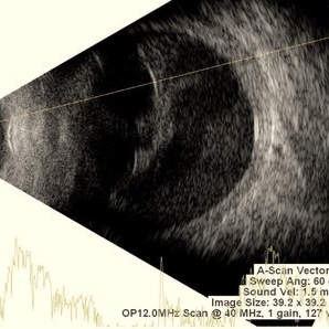
Membranous echoes with high spikes with restricted after movements suggestive of retinal detachment ( closed funnel configuration )
Photographer: Dr.Anand Temkar- Retina Foundation, Ahmedabad
Condition/keywords: A-scan ultrasound, B scan ultrasound, Closed funnel RD
-
 IOL-Drop
Oct 27 2023 by Anand Temkar
High spikes with restricted after movements suggestive of foreign body ( IOL ) in vitreous.
Photographer: Dr.Anand Temkar- Retina Foundation, Ahmedabad
Condition/keywords: A-scan ultrasound, B scan ultrasound, IOL drop
-
 CD
CD
Oct 27 2023 by Anand Temkar
Membranous echoes with moderate to high spikes with restricted after movements suggestive of choroidal detachment.
Photographer: Dr.Anand Temkar- Retina Foundation, Ahmedabad
Condition/keywords: A-scan ultrasound, B scan ultrasound, choroidal detachment
-
 Retinal Detachment
Oct 29 2023 by Anand Temkar
Membranous echoes with high spikes with restricted after movements suggestive of retinal detachmment.
Photographer: Dr.Anand Temkar- Retina Foundation, Ahmedabad
Condition/keywords: A-scan ultrasound, B scan ultrasound, open funnel RD
-
 Subhyaloid Haemorrhage
Oct 29 2023 by Anand Temkar
Multiple dot echoes with mild to moderate spikes with free after movements suggestive of subhyaloid haemorrhage.
Photographer: Dr.Anand Temkar- Retina Foundation, Ahmedabad
Condition/keywords: A-scan ultrasound, B scan ultrasound, subhyaloid blood, SUBHYALOID HEMORRHAGE
-
 Tractional retinal detachment
Oct 29 2023 by Anand Temkar
Membranous echoes with high spikes with restricted after movements suggestive of retinal detachment ( tractional ).
Photographer: Dr.Anand Temkar- Retina Foundation, Ahmedabad
Condition/keywords: A-scan ultrasound, B scan ultrasound, tractional retinal detachment
-
 Vitreous Haemorrhage
Oct 31 2023 by Anand Temkar
Multiple dot echoes with mild to moderate spikes with free aftermovements suggestive of vitreous haemorrhage
Photographer: Dr.Anand Temkar- Retina Foundation, Ahmedabad
Condition/keywords: A-scan ultrasound, B scan ultrasound, vitreous blood
-
 Vitreous degeneration
Oct 31 2023 by Anand Temkar
Few dot echoes with mild to moderate spikes with free aftermovements suggestive of vitreous degeneration
Photographer: Dr.Anand Temkar- Retina Foundation, Ahmedabad
Condition/keywords: A-scan ultrasound, B scan ultrasound
-
 Intraocular Foreign Body
Oct 31 2023 by Anand Temkar
Echoes with high spikes with restricted aftermovements suggestive od intraocular foreign body
Photographer: Dr.Anand Temkar- Retina Foundation, Ahmedabad
Condition/keywords: A-scan ultrasound, B scan ultrasound, intraocular foreign body
-
 Endophthalmitis
Nov 2 2023 by Anand Temkar
Multiple dot echoes with mild to moderate spikes with free after movements suggestive of vitreous exudates in a case of endophthalmitis.
Photographer: Dr.Anand Temkar- Retina Foundation, Ahmedabad
Condition/keywords: A-scan ultrasound, B scan ultrasound, endophthalmitis
-
 Choroidal Detachment
Nov 23 2023 by Anand Temkar
LE color photo montage showing choroidal detachment of a 63 years old male who gives history of LE filtration surgery with mitomycin c and anterior vitrectomy elsewhere a month ago.
Photographer: Dr.Anand Temkar- Retina Foundation, Ahmedabad
Imaging device: Mirante
Condition/keywords: choroidal detachment
-
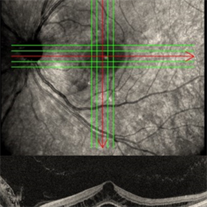
 Choroidal folds i/c/o hypotony
Nov 23 2023 by Anand Temkar
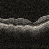
OCT showing choroidal folds in a follow up case of filtration surgery with mitomycin c and anterior vitrectomy elsewhere.
Photographer: Dr.Anand Temkar- Retina Foundation, Ahmedabad
Imaging device: Mirante
Condition/keywords: choroidal folds, hypotony, OCT
-
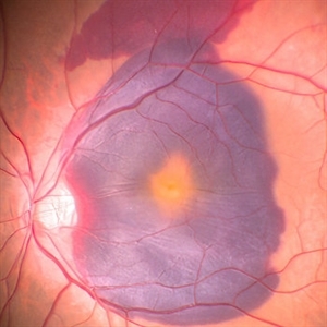
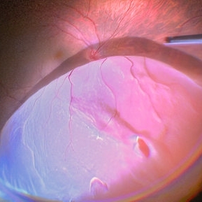
 Retinal detachment
Nov 23 2023 by Anand Temkar
LE color photo montage of a 50 years old male with supero-nasal retinal detachment (with break) and we can see horseshoe tear temporally with sub-retinal fluid.
Photographer: Dr.Anand Temkar- Retina Foundation, Ahmedabad
Imaging device: Mirante
Condition/keywords: RD, retinal break
-
 VH i/c/o Lasered PDR
May 8 2024 by Anand Temkar
Widefield fundus photograph of LE of a 58 years old male with Vitreous Hemorrhage inferiorly status post pan retinal photocogulation.
Photographer: Dr.Anand Temkar- Retina Foundation, Ahmedabad
Imaging device: Mirante
Condition/keywords: laser photocoagulation, proliferative diabetic retinopathy (PDR), vitreous hemorrhage
-
 Lasered Horse Shoe Tear with Mild Vitreous Haemorrhage
Jun 12 2024 by Anand Temkar
RE CF Montage of a 55 yrs old male with Lasered HST and Mild VH.
Photographer: Dr.Anand Temkar- Retina Foundation, Ahmedabad
Imaging device: Mirante
Condition/keywords: vitreous hemorrhage
-
 Nucleus in Vitreous
Jun 12 2024 by Anand Temkar
Intraopertive still of a 62 yrs old male with nucleus in vitreous ( LE ).
Photographer: Dr.Anand Temkar- Retina Foundation, Ahmedabad
Condition/keywords: lens drop
-
 Nucleus Drop
Jun 12 2024 by Anand Temkar
Intraoperative still of a 58 year old female with nucleus drop (LE).
Photographer: Dr.Anand Temkar- Retina Foundation, Ahmedabad
Condition/keywords: nucleus drop
-
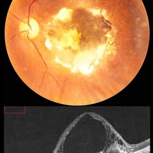
 Submacular Hemorrhage
Jun 12 2024 by Anand Temkar
Intraoperative still of a 58 year old male with submacular hemorrhage (LE).
Photographer: Dr.Anand Temkar- Retina Foundation, Ahmedabad
Condition/keywords: submacular hemorrhage, subretinal hemorrhage
-
 Dropped IOL on Retinal Surface (Over Macula)
Jun 12 2024 by Anand Temkar
Intra operative still of a 62 yrs old female with IOL drop (LE).
Photographer: Dr.Anand Temkar- Retina Foundation, Ahmedabad
Condition/keywords: IOL drop
-
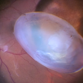
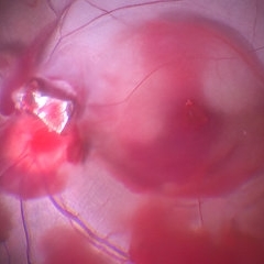
 Giant Retinal Tear with Choroidal Detachment
Jun 12 2024 by Anand Temkar
Intra operative still of a 34 year old male showing Giant Retinal Tear with Choroidal Detachment.
Photographer: Dr.Anand Temkar- Retina Foundation, Ahmedabad
Condition/keywords: choroidal detachment, giant retinal tear
-
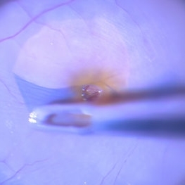
 Impacted Foreign Body on Retina
Jun 12 2024 by Anand Temkar
This is intra operative still of a impacted foreign body in the retina of a 28 year old male.
Photographer: Dr.Anand Temkar- Retina Foundation, Ahmedabad
Condition/keywords: intraocular foreign body
-
 ILM Peeling in a Case of Macular Hole
Jun 13 2024 by Anand Temkar
Intraoperative still of a 62 year old female with macular hole.
Photographer: Dr.Anand Temkar- Retina Foundation, Ahmedabad
Condition/keywords: ILM peeling, macular hole
-
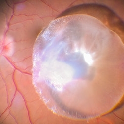
 Giant Retinal Tear With Retinal Fold
Jun 13 2024 by Anand Temkar
Intraoperative still of a 34 year old male showing giant retinal tear with retinal fold.
Photographer: Dr.Anand Temkar- Retina Foundation, Ahmedabad
Condition/keywords: giant retinal tear, GRT, retinal fold
-
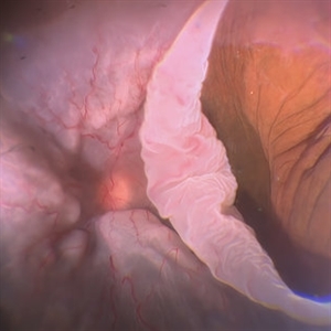
 Superior RD with HST Superiorly
Jun 13 2024 by Anand Temkar
Intraoperative still of a 48 year old male showing RD with superior HST and a break.
Photographer: Dr.Anand Temkar- Retina Foundation, Ahmedabad
Condition/keywords: RD
-
 IOL Drop
Jun 13 2024 by Anand Temkar
Intraoperative still of a 72 year old male showing dropped IOL.
Photographer: Dr.Anand Temkar- Retina Foundation, Ahmedabad
Condition/keywords: IOL drop
-
 Metallic Foreign Body Removed From Site of Impaction Intra Operatively
Jun 13 2024 by Anand Temkar
Intraoperative still of a 27 year old male showing metallic foreign body removed from site of impaction from retina.
Photographer: Dr.Anand Temkar- Retina Foundation, Ahmedabad
Condition/keywords: metallic foreign body
-
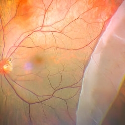
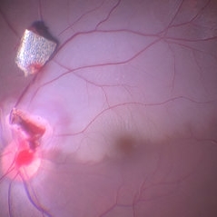
 Left Eye Optical Coherence Tomography Showing Optic Disc Pit
Nov 9 2024 by Anand Temkar
Left Eye Optical Coherence Tomography of a 48 years old male patient showing Optic Disc Pit.
Photographer: Dr.Anand Temkar- Retina Foundation, Ahmedabad
Imaging device: Mirante
Condition/keywords: optic disc pit, Optic pit
-
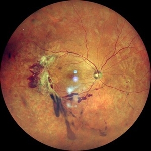
 Macular Mount Everest
Aug 8 2025 by Anand Temkar
A 75 yrs old male came with the chief complains of DOV in LE since past 20 yrs. His BCVA in RE was 6/9 and in LE, it was CF 1 meter. His IOP was 13 mm of Hg in RE and 15 mm of Hg in LE. Patient is a k/c/o DM type 2 since past 20 yrs and is on regular medication. Patient is a k/c/o solitary kidney. Patient gives h/o ( LE ) Intravitreal injection Avastin 3 times 13 yrs ago i/c/o CNVM. In the LE color photo we can see the scarred CNVM along with altered foveal contour. LE OCT also shows cystic spaces with large elevation and scarring.
Photographer: Dr.Anand Temkar- Vasan Eye Hospital, Tiruchirapalli
Condition/keywords: CNVM, macular edema, scarred cnvm
A project from the American Society of Retina Specialists