-
 Coats disease
Nov 8 2022 by Heitor Nogueira
Fundus photograph of an 12-year-old asymptomatic patient. It is possible to observe the presence of vascular telangiectasias associated with areas of exudation without the presence of a tumor lesion.
Photographer: Heitor Nogueira, Instituto Penido Burnier, Campinas-SP, Brazil
Condition/keywords: Coats' disease
-
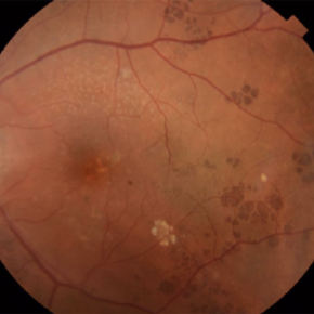 CHRPE - "BEAR TRACKS" PATTERN
Nov 8 2022 by Heitor Nogueira
BEAR TRACKS
Photographer: Heitor Nogueira
Condition/keywords: bear tracks, CHRPE, congenital hypertrophy of the retinal pigment epithelium (CHRPE)
-
Spontaneous lens dislocation (Weill Marchesani Syndrome)
Nov 9 2022 by Heitor Nogueira
A 9 year-old Male patient diagnosed with Weill Marchesani presented spontaneous bilateral lens dislocation. Weill-Marchesani syndrome, also known as spherophakia-brachymorphy syndrome and mesodermal dysmorphodystrophy, is an inherited connective tissue disorder characterized by eye lens abnormalities, secondary glaucoma, short stature, brachydactyly, joint stiffness, and cardiovascular defects.
Photographer: Heitor Nogueira, Insituto Penido Burnier, Campinas-SP, Brazil
Condition/keywords: mesodermal dysmorphodystrophy, spherophakia-brachymorphy syndrome, spontaneous lens dislocation, video, Weill Marchesani Syndrome
-
 Extensive Macular Atrophy with Pseudodrusen (EMAP)
Jul 2 2023 by Heitor Nogueira
Fundus photograph of an 56-year-old woman with a macular atrophy caused by EMAP. We can also observe the presence of Paving stones grouped throughout the middle and extreme periphery.
Photographer: Heitor Nogueira, Instituto Penido Burnier, Campinas, São Paulo, Brazil.
Imaging device: Optos California
Condition/keywords: macular atrophy, paving stone degeneration
-
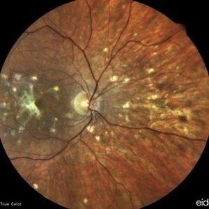
 Punctate inner choroidopathy (PIC) with CNVM.
Oct 18 2023 by Heitor Nogueira
Fundus photograph of a 29-year-old woman with a 2-week history of low visual acuity associated with central scotoma. Ophthalmological history of axial myopia of -4,00D. She denied personal and family history.
Photographer: Heitor Nogueira, Instituto Penido Burnier, Campinas-SP, Brazil.
Imaging device: Eidon True Color
Condition/keywords: choroidal neovascularization (CNV), CNVM, multifocal chorioretinitis (MCP), punctate inner choroidopathy (PIC)
-
 Punctate inner choroidopathy (PIC) with CNVM
Oct 18 2023 by Heitor Nogueira
Fundus photograph of a 29-year-old woman with a 2-week history of low visual acuity associated with central scotoma. Ophthalmological history of axial myopia of -4,00D. She denied personal and family history.
Photographer: Heitor Nogueira, Instituto Penido Burnier, Campinas-SP, Brazil.
Imaging device: Eidon True Color
Condition/keywords: CNVM, multifocal chorioretinitis (MCP), punctate inner choroidopathy (PIC)
-
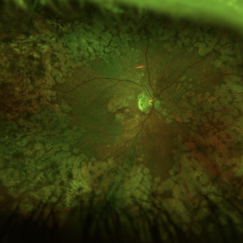
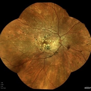 Idiopathic Multifocal Choroiditis
Oct 25 2023 by Heitor Nogueira
Fundus photograph of a 48-year-old woman with idiopathic multifocal choroiditis
Photographer: Heitor Nogueira, Instituto Penido Burnier, Campinas-SP, Brazil
Imaging device: Eidon
Condition/keywords: multifocal choroiditis, multifocal hypofluorescent lesions
-
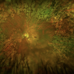
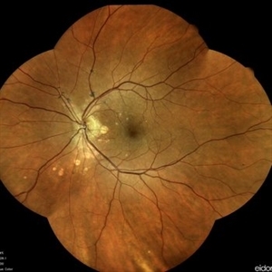 Idiopathic Multifocal Choroiditis
Oct 25 2023 by Heitor Nogueira
Fundus photograph of a 48-year-old woman with idiopathic multifocal choroiditis.
Photographer: Heitor Nogueira, Instituto Penido Burnier, Campinas-SP, Brazil
Imaging device: Eidon
Condition/keywords: choroiditis
-
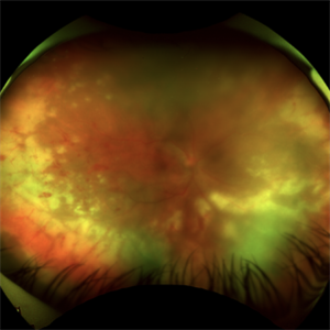
 Acute Retinal Necrosis
Jul 3 2025 by Heitor Nogueira
Fundus photograph of an 53-year-old woman with patient who reported unilateral visual acuity loss for 10 days associated with ocular pain. She presented conjunctival hyperemia with temporal and nasal nodular scleritis, anterior chamber reaction 2+/4+, Koeppe nodules, granulomatous PKs, vitritis 2+/4+, multiple areas of vasculitis in arcades and periphery, associated with hemorrhages and necrotizing retinitis in temporal, inferior and nasal periphery. patient who reported unilateral visual acuity loss for 10 days associated with ocular pain. He presented conjunctival hyperemia with temporal and nasal nodular scleritis, anterior chamber reaction 2+/4+, Koeppe nodules, granulomatous PKs, vitreitis 2+/4+, multiple areas of vasculitis in the arcades and periphery, associated with hemorrhages and necrotizing retinitis in the temporal, inferior and nasal periphery. Positive serology for Herpes Virus.
Photographer: Heitor Nogueira, Penido Burnier Institute and CHOV, Campinas, São Paulo, Brazil
Imaging device: Optos Daytona
Condition/keywords: ARN complications, Herpes, progressive outer retinal necrosis (PORN)
-
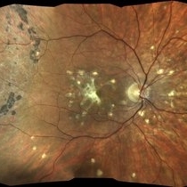
 Acute Retinal Necrosis (ARN)
Jul 3 2025 by Heitor Nogueira
Fundus photograph of an 63-year-old woman who reported unilateral visual acuity loss for 10 days associated with ocular pain. He presented conjunctival hyperemia with temporal and nasal nodular scleritis, anterior chamber reaction 2+/4+, Koeppe nodules, granulomatous PKs, vitreitis 2+/4+, multiple areas of vasculitis in the arcades and periphery, associated with hemorrhages and necrotizing retinitis in the temporal, inferior and nasal periphery. Positive serology for Herpes Virus
Photographer: Heitor Nogueira, Penido Burnier Institute, Campinas, São Paulo, Brazil
Imaging device: Optos Daytona
Condition/keywords: ARN complications, Herpes, progressive outer retinal necrosis (PORN), Uveitis
A project from the American Society of Retina Specialists