-
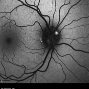
 Blue autofluroscence of Left eye optic nerve head showing auto fluorescence of the drusen
Aug 5 2022 by Kavitha Duraipandi, MD DNB FICO FRCS
Blue autofluroscence of Right eye optic nerve head showing auto fluorescence of the drusen
Photographer: Natalie Fox- Bussell
Condition/keywords: Autoflourescence, Heidelburg Spectralis
-
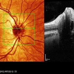
 Left eye SD- OCT-RNFL of optic nerve head drusen showing hypo reflective centre with hyper reflective margins.
Aug 5 2022 by Kavitha Duraipandi, MD DNB FICO FRCS
A 20 year old patient referred to the clinic with blurred disc margins to rule out papilledema.
Photographer: Natalie Fox- Bussell
Condition/keywords: optic nerve drusen, optical coherence tomography (OCT)
-
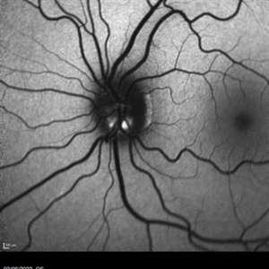
 Blue autofluroscence of Right eye optic nerve head showing auto fluorescence of the drusen
Aug 5 2022 by Kavitha Duraipandi, MD DNB FICO FRCS
A 20 year old patient referred to the clinic with blurred disc margins to rule out papilledema.
Photographer: Natalie Fox- Bussell
Condition/keywords: Blue autofluroscence, Heidelburg Spectralis
-
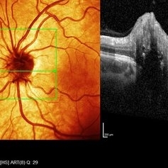
 Right eye SD- OCT-RNFL of optic nerve head drusen showing hypo reflective centre with hyper reflective margins.
Aug 5 2022 by Kavitha Duraipandi, MD DNB FICO FRCS
A 20 year old patient referred to the clinic with blurred disc margins to rule out papilledema.
Photographer: Natalie Fox- Bussell
Condition/keywords: optic nerve drusen, optical coherence tomography (OCT)
-
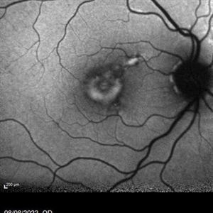 Adult onset foveomacular vitelliform dystrophy- Blue autofluroscence
Aug 10 2022 by Kavitha Duraipandi, MD DNB FICO FRCS
Blue autofluroscence in a 80 year old patient showing increased autofluroscence in the vitelliform lesion.
Photographer: Paul Cunning, Blackpool Victoria Hospital
Condition/keywords: aovd
-
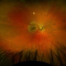 CHRPE
CHRPE
Jan 6 2025 by Kavitha Duraipandi, MD DNB FICO FRCS
Bear tracks (animal tracks, grouped pigmentation spots) are simply many small CHRPEs located in isolated small area of the retina. These have not been reported to have the potential to transform into adenocarcinoma but yearly evaluations may be prudent.
Condition/keywords: CHRPE
-
OCT Video Imaging of Left Eye Age Related Macular Degeneration
Jan 6 2025 by Kavitha Duraipandi, MD DNB FICO FRCS
Left eye OCT macula shows various biomarkers like PED, sub retinal fluid, sub retinal hyper reflective material and hyper reflective foci suggestive of wet age-related macular degeneration.
Condition/keywords: OCT biomarkers, wet age-related macular degeneration (wet AMD)
A project from the American Society of Retina Specialists