-
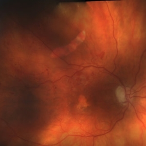 Extrafoveal PED with RPE rip colour photo
Dec 23 2012 by Alex P. Hunyor, MD
80-year-old female with subfoveal occult CNV and large extrafoveal PED which underwent spontaneous RPE rip.
Condition/keywords: pigment epithelial detachment (PED), retinal pigment epithelium (RPE) tear
-
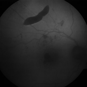 Extrafoveal PED with RPE rip AF
Dec 23 2012 by Alex P. Hunyor, MD
80-year-old female with subfoveal occult CNV and large extrafoveal PED which underwent spontaneous RPE rip. Autofluorescence image shows hypoautofluorescence in crescentic area of absent RPE due to rip, and also RPE atrophy adjacent to fovea. Intervening small areas of hypoautofluorescence are due to subretinal haemorrhage.
Condition/keywords: pigment epithelial detachment (PED), retinal pigment epithelium (RPE) tear
-
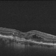 RPE rip macular OCT
Dec 23 2012 by Alex P. Hunyor, MD
80-year-old female with subfoveal occult CNV and large extrafoveal PED which underwent spontaneous RPE rip. OCT shows subfoveal CNV and intraretinal cystic edema
Condition/keywords: pigment epithelial detachment (PED), retinal pigment epithelium (RPE) tear
-
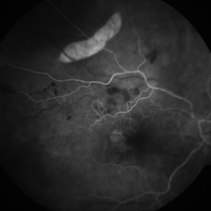 Extrafoveal PED with RPE rip FA1
Dec 23 2012 by Alex P. Hunyor, MD
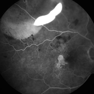
80-year-old female with subfoveal occult CNV and large extrafoveal PED which underwent spontaneous RPE rip. Early phase FA showing intense hyperfluorescence in the area of acute absence of RPE.
Condition/keywords: pigment epithelial detachment (PED), retinal pigment epithelium (RPE) tear
-
 Extrafoveal PED with RPE rip FA2
Dec 23 2012 by Alex P. Hunyor, MD
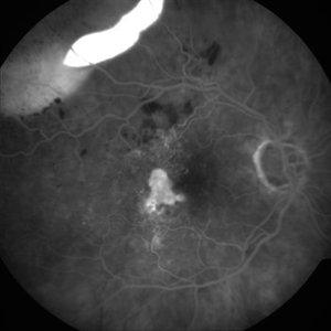
80-year-old female with subfoveal occult CNV and large extrafoveal PED which underwent spontaneous RPE rip. FA shows intense hyperfluorescence in area of absent RPE, progressive filling of extrafoveal PED, and hyperfluorescence in macula from atrophy and occult CNV.
Condition/keywords: pigment epithelial detachment (PED), retinal pigment epithelium (RPE) tear
-
 Extrafoveal PED with RPE rip FA3
Dec 23 2012 by Alex P. Hunyor, MD
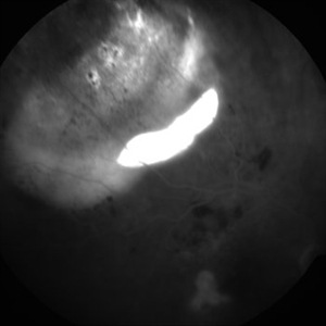
80-year-old female with subfoveal occult CNV and large extrafoveal PED which underwent spontaneous RPE rip. FA shows intense hyperfluorescence in area of absent RPE, progressive filling of extrafoveal PED, and hyperfluorescence in macula from atrophy and occult CNV.
Condition/keywords: pigment epithelial detachment (PED), retinal pigment epithelium (RPE) tear
-
 Extrafoveal PED with RPE rip FA4
Dec 23 2012 by Alex P. Hunyor, MD
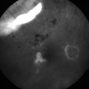
80-year-old female with subfoveal occult CNV and large extrafoveal PED which underwent spontaneous RPE rip. FA shows intense hyperfluorescence in area of absent RPE, progressive filling of extrafoveal PED, and hyperfluorescence in macula from atrophy and occult CNV.
Condition/keywords: pigment epithelial detachment (PED), retinal pigment epithelium (RPE) tear
-
 Extrafoveal PED with RPE rip FA5
Dec 23 2012 by Alex P. Hunyor, MD
80-year-old female with subfoveal occult CNV and large extrafoveal PED which underwent spontaneous RPE rip. FA shows intense hyperfluorescence in area of absent RPE, progressive filling of extrafoveal PED, and hyperfluorescence in macula from atrophy and occult CNV.
Condition/keywords: pigment epithelial detachment (PED), retinal pigment epithelium (RPE) tear
A project from the American Society of Retina Specialists