-
 Posterior Scleral Laceration
May 24 2022 by Ahmad B. Tarabishy, MD
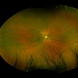
A 49 year old male was referred from the ER following an injury to his right medial eyelid with a sharp metal tip. He had brief pain at the time. No new floaters, flashes, or blurred vision. Intraocular pressure was 18 OS. Examination showed a full thickness laceration of the nasal posterior globe with adjacent hemorrhage. Prophylactic laser coagulation was performed. Examination 2 weeks later shows maturing laser scars and no complications related to the scleral laceration. The patient reports no new vision changes.
Photographer: Angelo Rico MD, Retina Specialists of Tampa
Imaging device: Optos
Condition/keywords: globe perforation, scleral laceration
-
 Posterior Scleral Laceration
May 24 2022 by Ahmad B. Tarabishy, MD
A 49 year old male was referred from the ER following an injury to his right medial eyelid with a sharp metal tip. He had brief pain at the time. No new floaters, flashes, or blurred vision. Intraocular pressure was 18 OS. Examination showed a full thickness laceration of the nasal posterior globe with adjacent hemorrhage. Prophylactic laser coagulation was performed. Examination 2 weeks later shows maturing laser scars and no complications related to the scleral laceration. The patient reports no new vision changes.
Photographer: Angelo Rico MD, Retina Specialists of Tampa
Imaging device: Optos
Condition/keywords: corneal laceration, globe perforation
A project from the American Society of Retina Specialists