-
 Proliferative Diabetic Retinopathy
Sep 16 2021 by Tandava Krishnan
Fundus photograph of a 60 year old poorly controlled diabetic who presented with decreased vision in left eye.
Photographer: Tandava Krishnan
Condition/keywords: fibrovascular proliferation, neovascularization elsewhere (NVE), proliferative diabetic retinopathy (PDR), subhyaloid hemorrhage
-
 Sub Hyaloid Haemorrhage after Valsalva manoeuvre
Jun 29 2022 by Tandava Krishnan
Left eye fundus picture of a 35 year old gentleman showing sub hyaloid haemorrhage after Valsalva manoeuvre
Photographer: Dr Tandava Krishnan, Dr Agarwal's Eye Hospital, Hyderabad, India
Condition/keywords: Green LASER
-
 Sub hyaloid haemorrhage 10 minutes after LASER hyaloidotomy
Jun 29 2022 by Tandava Krishnan
This is the left eye fundus picture of a 35 year old man, 10 minutes after Green LASER Hyaloidotomy showing clearing of blood from the sub hyaloid space into the vitreous
Photographer: Dr Tandava Krishnan, Dr Agarwal's eye Hospital, Hyderabad, India
Condition/keywords: Green LASER
-
 10 days after Green LASER hyaloidotomy
Jun 29 2022 by Tandava Krishnan
This is the left eye fundus picture of a 35 year old gentleman 10 days after LASER hyaloidotomy
Photographer: Dr Tandava Krishnan, Dr Agarwal's eye Hospital , Hyderabad, India
Condition/keywords: Green LASER
-
 Branch Retinal Artery occlusion
Sep 3 2022 by Tandava Krishnan
Branch Retinal artery occlusion with a visible y shaped embolus at the point of division of arteriole
Photographer: Tandava Krishnan
Condition/keywords: branch retinal artery occlusion (BRAO), embolic, embolus
-
 Myelinated nerve fibre
Nov 10 2022 by Tandava Krishnan
Red free fundus photograph of the left eye of a patient with myelinated nerve fibre. Myelinated nerve fibres are autofluorescent and can be clearly made out on red free imaging
Condition/keywords: myelinated nerve fibers
-
 Myelinated Nerve Fiber
Nov 10 2022 by Tandava Krishnan
Color fundus photo of the Left eye of a patient showing myelinated nerve fiber
Condition/keywords: myelinated nerve fibers
-
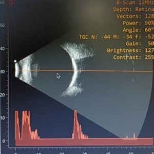 Choroidal Osteoma
Nov 10 2022 by Tandava Krishnan
B scan imaging of the eye with choroidal osteoma showing high reflective choroidal lesion with Posterior shadowing suggestive of a bone like lesion
Condition/keywords: choroidal osteoma, choroidal tumor, choroidal tumour, macular choroidal osteoma
-
 Choroidal osteoma
Nov 10 2022 by Tandava Krishnan
Right eye fundus picture of a patient with Creamy yellow choroidal lesion suggestive of Choroidal osteoma
Condition/keywords: choroidal osteoma, choroidal tumor, choroidal tumour, macular choroidal osteoma
A project from the American Society of Retina Specialists