-
 Stargardt's disease
Aug 18 2021 by Priyanka Raj, MBBS, MS
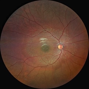
Fundus photograph of a 24 year-old male with Stargardt's disease.
Photographer: Priyanka Raj, Prakash Netra Kendr, Lucknow, India
Imaging device: Zeiss Clarus 500
Condition/keywords: fundus flavimaculatus, heredomacular degeneration, Stargardt disease
-
 Stargardt's disease
Aug 18 2021 by Priyanka Raj, MBBS, MS
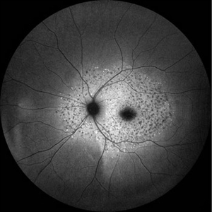
Fundus photograph of a 24 year-old male with Stargardt's disease
Photographer: Priyanka Raj, Prakash Netra Kendr, Lucknow, India
Imaging device: Zeiss Clarus 500
Condition/keywords: fundus flavimaculatus, heredomacular degeneration, Stargardt disease
-
 Stargardt's disease
Aug 18 2021 by Priyanka Raj, MBBS, MS
Fundus photograph of a 24 year-old male with Stargardt's disease
Photographer: Priyanka Raj, Prakash Netra Kendr, Lucknow, India
Imaging device: Zeiss Clarus 500
Condition/keywords: fundus flavimaculatus, Stargardt disease
-
 Stargardt's Disease
Aug 18 2021 by Priyanka Raj, MBBS, MS
Fundus photograph of a 24 year-old male with Stargardt's disease
Photographer: Priyanka Raj, Prakash Netra Kendr, Lucknow, India
Imaging device: Zeiss Clarus 500
Condition/keywords: fundus flavimaculatus, heredomacular degeneration, Stargardt disease
-
 Sneaky bubble under the buckle
May 19 2022 by Priyanka Raj, MBBS, MS
First post-operative day picture of a highly myopic eye of a patient who underwent superior 120 degrees 276 circumferential scleral buckle for bullous retinal detachment. The picture shows a remnant air bubble used as a tamponade, restricted by the buckle height and a break treated with cryopexy, well supported by the buckle.
Photographer: Ajeet, Prakash Netra Kendra
Imaging device: Zeiss Clarus 500
Condition/keywords: Cryopexy, high myopia, myopic fundus, retinal tear, scleral buckle
-
 Retinal angiomatous proliferation (RAP)
Jun 15 2022 by Priyanka Raj, MBBS, MS
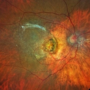
Retinochoroidal anastomosis seen in stage III Retinal angiomatous proliferation (RAP).
Photographer: Sushil Mishra
Imaging device: Zeiss Clarus 500
Condition/keywords: age-related macular degeneration (AMD), retinal angiomatous proliferation (RAP), retinochoroidal anastomosis
A project from the American Society of Retina Specialists