-
 Ischemic CRVO
Jun 23 2021 by Eduardo Torres-Porras, MD
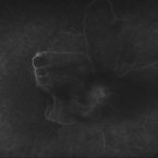
Retinal fluorangiography of the right eye of a 36-year-old male who had an ischemic CRVO following a hypertensive emergency secondary to consumption of high doses of cocaine. The image shows marked areas of ischemia of the retina with only perfusion of the macular area. In the nasal mid-to-far peripheral retina, NVs can be observed.
Photographer: Eduardo Torres-Porras, Provissia, Laser y Ultrasonido Ocular
Imaging device: Optos, Californina
Condition/keywords: central retinal vein occlusion (CRVO), ischemic CRVO, optical coherence tomography (OCT), ultra-wide field imaging
A project from the American Society of Retina Specialists