-
 Retinal Emboli and Resolution
Jun 17 2021 by Jonathan S Chang, MD, FASRS
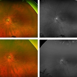
This patient developed a cerebral vascular accident and underwent neuroendovascular thrombectomy. Following the thrombectomy the patient noted transient blurred vision. During that time the patient was also noted to have significant left carotid artery disease. Top row widefield photos are color (left) and red free (right) of left eye fundus showing small scattered emboli, especially superonasally, inferiorly and inferotemporally. Bottom row color (left) and red free (right) widefield photos taken five months after presentation, showing resolution of some of the emboli, but persistence of the superonasal embolus.
Condition/keywords: embolus, retinal arteriolar occlusion
A project from the American Society of Retina Specialists