-
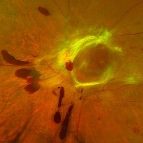 Wolf Jaw
Jun 23 2019 by Veronika Yehezkeli
Wolf Jaw
Imaging device: Optos
Condition/keywords: ischemia, neovascularization (NV), neovascularization elsewhere (NVE), preretinal hemorrhage, proliferative diabetic retinopathy (PDR), wolf jaw
-
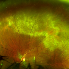 Massive Commotio Retinae
Oct 20 2020 by Veronika Yehezkeli
24-year-old man was injured from an explosion of a plastic bottle towards the nasal conjunctiva of his left eye. A massive commotio retinae was diagnosed superotemporally.
Photographer: Veronika Yehezkeli, Meir medical center, Israel
Condition/keywords: blunt trauma, commotio retinae, preretinal hemorrhage
-
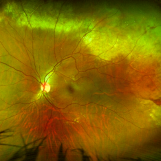 Massive Commotio Retinae
Oct 20 2020 by Veronika Yehezkeli
Fundus photograph of a 24-year-old male, made after blunt trauma with a plastic bottle. Note massive commotio retinae and preretinal hemorrhages in the contralateral to trauma area.
Photographer: Veronika Yehezkeli, Meir medical center, Israel
Condition/keywords: blunt trauma, commotio retinae, preretinal hemorrhage
-
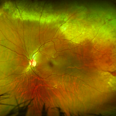 Massive Commotio Retinae
Oct 20 2020 by Veronika Yehezkeli
Fundus photograph of a 24-year-old male, made after blunt trauma with a plastic bottle. Note massive commotio retinae and preretinal hemorrhages in the contralateral to trauma area.
Photographer: Veronika Yehezkeli, Meir medical center, Israel
Condition/keywords: blunt trauma, commotio retinae, preretinal hemorrhage, trauma
-
 Regressed ROP with Retinal Vascularization Extending Beyond the Zone II Ridge
Oct 25 2026 by Veronika Yehezkeli
Fundus photograph of the right eye of a 12-year-old girl with regressed retinopathy of prematurity, treated in infancy with intravitreal Bevacizumab. A fibrotic regressed ridge is seen in zone II temporally, with retinal vessels extending beyond the ridge into the previously avascular peripheral retina. Superiorly (12–1 o’clock), an area of vitreoretinal traction containing retinal tears and atrophic holes has just been treated with barrier laser retinopexy, seen as fresh laser spots. Old laser scars are present superotemporally. The image serves as a baseline for long-term follow-up and illustrates the late peripheral vitreoretinal complications that can develop years after ROP regression.
Photographer: Patricia Goldberg
Condition/keywords: intravitreal bevacizumab, REGRESSED ROP, retinopathy of prematurity (ROP), white without pressure

A project from the American Society of Retina Specialists