-
 Ocular Histoplasmosis
Mar 27 2019 by Gary R. Cook, MD, FACS
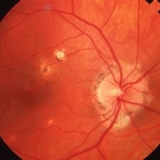
Fellow eye (OD) of a 41-year-old white female with ocular histoplasmosis showing peripapillary atrophy and several atrophic histo spots OD; no CNVM present; V.A.= 20/20.
Imaging device: Topcon VT-50
Condition/keywords: atrophic spot, histoplasmosis, peripapillary atrophy, presumed ocular histoplasmosis syndrome (POHS)
-
 Histo and Subfoveal Neovascular Membrane
Mar 27 2019 by Gary R. Cook, MD, FACS
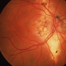
41-year-old white female with a large subfoveal CNVM, subretinal fluid, and hemorrhage secondary to presumed ocular histoplasmosis (POHS) OS; V.A.= 20/400.
Imaging device: Topcon VT-50
Condition/keywords: hemorrhage, peripapillary atrophy, presumed ocular histoplasmosis syndrome (POHS), subfoveal choroidal neovascularization, subfoveal neovascular membrane
-
 Histoplasmosis with Choroidal Neovascularization
Mar 27 2019 by Gary R. Cook, MD, FACS
59-year-old white male with presumed ocular histoplasmosis (POHS) and a choroidal neovascular membrane (CNVM) along the temporal margins of the peripapillary atrophy; V.A.= 20/80+2.
Imaging device: Topcon VT-50
Condition/keywords: choroidal neovascular membrane (CNVM), peripapillary atrophy, presumed ocular histoplasmosis syndrome (POHS)
-
 Histoplasmosis and Subfoveal Neovascular Membrane
Mar 27 2019 by Gary R. Cook, MD, FACS
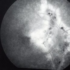
Mid-phase (20.4 seconds) fluorescein angiogram image of the right eye of 59-year-old white male with ocular histoplasmosis and a well-defined subfoveal CNVM OD; V.A.= 20/80+2
Imaging device: Topcon VT-50
Condition/keywords: FA mid phase, fluorescein angiogram (FA), ocular histoplasmosis syndrome (OHS), peripapillary atrophy, presumed ocular histoplasmosis syndrome (POHS), subfoveal choroidal neovascularization, subfoveal neovascular membrane
-
 Histoplasmosis and Subfoveal Neovascular Membrane
Mar 27 2019 by Gary R. Cook, MD, FACS
Late-phase fluorescein angiogram image of the right eye of a 59-year-old white male with ocular histoplasmosis and a subfoveal neovascular membrane showing late leakage and diffusion of dye from the membrane; V.A.= 20/80+2.
Imaging device: Topcon VT-50
Condition/keywords: FA late phase, fluorescein angiogram (FA), ocular histoplasmosis syndrome (OHS), peripapillary atrophy, presumed ocular histoplasmosis syndrome (POHS), subfoveal neovascular membrane
-
 Histoplasmosis and Old Disciform Macular Scar
Mar 27 2019 by Gary R. Cook, MD, FACS
Left eye of a 59-year-old white male with an old, inactive, disciform macular scar secondary to presumed ocular histoplasmosis (POHS); V.A.= counting fingers at 3 feet.
Imaging device: Topcon VT-50
Condition/keywords: central disciform scar, disciform scar, peripapillary atrophy, presumed ocular histoplasmosis syndrome (POHS)
-
 Histoplasmosis and Subfoveal Neovascular Membrane
Mar 27 2019 by Gary R. Cook, MD, FACS
26-year-old white female with new-onset subfoveal CNVM secondary to presumed ocular histoplasmosis (POHS) in her right eye; V.A.= 20/200.
Imaging device: Topcon VT-50
Condition/keywords: presumed ocular histoplasmosis syndrome (POHS), subfoveal choroidal neovascularization, subfoveal neovascular membrane
A project from the American Society of Retina Specialists