-
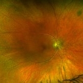 Vitamin A Retinopathy - Initial Photo OD
May 27 2019 by John S. King, MD
45-year-old female with a history of gastric bypass surgery, who was referred to Dr. Zocchi for as a possible choroidal dystrophy; patient had severe nyctalopia that had progressed over a year; acuity was 20/20 OU with normal IOP and A/C findings. The posterior segment showed multiple yellow-white punctate dots in the mid-periphery and periphery (see photo). Findings were consistent with probable vitamin A deficiency. Patient is getting vitamin levels checked by PCP and started on vitamin A, and will be seen back in a month.
Photographer: Shelly Blair
Imaging device: Optos CA
Condition/keywords: fleck retinopathy, malabsorption, nyctalopia, vitamin A deficiency, xerophthalmia
-
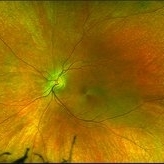 Vitamin A Retinopathy - Initial Photo OS
May 27 2019 by John S. King, MD
45-year-old female with a history of gastric bypass surgery, who was referred to Dr. Zocchi for as a possible choroidal dystrophy; patient had severe nyctalopia that had progressed over a year; acuity was 20/20 OU with normal IOP and A/C findings. The posterior segment showed multiple yellow-white punctate dots in the mid-periphery and periphery (see photo). Findings were consistent with probable vitamin A deficiency. Patient was started on Vit A, and levels were found to be very low. Follow up is in a month.
Photographer: Shelly Blair
Imaging device: Optos CA
Condition/keywords: fleck retinopathy, malabsorption, nyctalopia, vitamin A deficiency, xerophthalmia
-
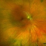 Vitamin A Deficiency Retinopathy Few Months Post Treatment With Resolution of the Retinopathy OD
Nov 17 2019 by John S. King, MD
45-year-old female with a history of gastric bypass surgery, who was referred to Dr. Zocchi for as a possible choroidal dystrophy; patient had severe nyctalopia that had progressed over a year; acuity was 20/20 OU with normal IOP and A/C findings. The posterior segment showed multiple yellow-white punctate dots in the mid-periphery and periphery (see initial photo). Findings were consistent with probable vitamin A deficiency. Vitamin A levels in the serum were greatly reduced. Patient was started on Vit A weekly infusions and PO daily. A few months later the punctate spots had resolved (there are some drusen like deposits in far periphery) and her nyctalopia had reversed.
Photographer: Brandon Peter
Imaging device: Optos CA
Condition/keywords: fleck retinopathy, malabsorption, nyctalopia, vitamin A deficiency, xerophthalmia
-
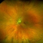 Vitamin A Deficiency Retinopathy Few Months Post Treatment With Resolution of the Retinopathy OS
Nov 17 2019 by John S. King, MD
45-year-old female with a history of gastric bypass surgery, who was referred to Dr. Zocchi for as a possible choroidal dystrophy; patient had severe nyctalopia that had progressed over a year; acuity was 20/20 OU with normal IOP and A/C findings. The posterior segment showed multiple yellow-white punctate dots in the mid-periphery and periphery (see initial photo). Findings were consistent with probable vitamin A deficiency. Patient was started on Vit A, and levels were found to be very low. Vitamin A levels in the serum were greatly reduced. Patient was started on Vit A weekly infusions and PO daily. A few months later the punctate spots had resolved (there are some drusen like deposits in far periphery) and her nyctalopia had reversed.
Photographer: Brandon Peter
Imaging device: Optos CA
Condition/keywords: fleck retinopathy, malabsorption, nyctalopia, vitamin A deficiency, xerophthalmia
-
 Vitamin A Deficiency Retinopathy - Initial Images on the Right and 5 Months Later on the Left
Nov 17 2019 by John S. King, MD
45-year-old female with a history of gastric bypass surgery, who was referred to Dr. Zocchi for as a possible choroidal dystrophy; patient had severe nyctalopia that had progressed over a year; acuity was 20/20 OU with normal IOP and A/C findings. The posterior segment showed multiple yellow-white punctate dots in the mid-periphery and periphery (see photo). Findings were consistent with probable vitamin A deficiency. Vitamin A levels in the serum were greatly reduced. Patient was started on Vit A weekly infusions and PO daily. A few months later the punctate spots had resolved (there are some drusen like deposits in far periphery) and her nyctalopia had reversed.
Photographer: Brandon Peter (L Image) and Shelly Blair (R Image)
Imaging device: Optos CA
Condition/keywords: fleck retinopathy, malabsorption, nyctalopia, vitamin A deficiency, xerophthalmia
-
 Vitamin A Deficiency Retinopathy - Initial Images - Late FA (R) and AF (L)
Nov 17 2019 by John S. King, MD
45-year-old female with a history of gastric bypass surgery, who was referred to Dr. Zocchi for as a possible choroidal dystrophy; patient had severe nyctalopia that had progressed over a year; acuity was 20/20 OU with normal IOP and A/C findings. The posterior segment showed multiple yellow-white punctate dots in the mid-periphery and periphery (see photo). Findings were consistent with probable vitamin A deficiency. Patient was started on Vit A, and levels were found to be very low. This is the initial images and the punctate spots are not detectable on the FA (no staining) or AF.
Photographer: Shelly Blair
Imaging device: Optos CA
Condition/keywords: fleck retinopathy, malabsorption, nyctalopia, vitamin A deficiency, xerophthalmia
A project from the American Society of Retina Specialists