-
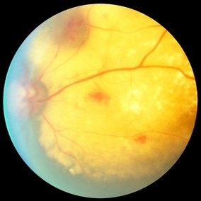 Coats Disease Slide 1
Oct 22 2012 by Ronald C. Gentile, MD
A unilateral, sub-retinal, and yellowish exudative lesion with associated retinal telangiectasias involving the nasal retina. Refractile elements can be seen and represent cholesterol crystals.
Photographer: The New York Eye & Ear Infirmary Department of Medical Imaging
Condition/keywords: congenital retinal telangiectasis
-
 Coats Disease Slide 2
Oct 22 2012 by Ronald C. Gentile, MD
Telangiectatic vessels are seen above the subretinal exudation. The abnormal vessels are tortuous and dilated.
Photographer: The New York Eye & Ear Infirmary Department of Medical Imaging
Condition/keywords: congenital retinal telangiectasis
-
 Coats Disease Slide 3
Oct 22 2012 by Ronald C. Gentile, MD
Flourescein angiogram with early filling of telangectatic vessels.
Photographer: The New York Eye & Ear Infirmary Department of Medical Imaging
Condition/keywords: congenital retinal telangiectasis
-
 Coats Disease Slide 4
Oct 22 2012 by Ronald C. Gentile, MD
Flourescein angiogram with progressive filling of telangectatic vessels. The abnormal vessels become more prominent and the dilated aneurysmal-like vessels fill.
Photographer: The New York Eye & Ear Infirmary Department of Medical Imaging
Condition/keywords: congenital retinal telangiectasis
-
 Coats Disease Slide 5
Oct 22 2012 by Ronald C. Gentile, MD
Flourescein angiogram with leakage of the telangectatic and dilated aneurysmal-like abnormal vessels.
Photographer: The New York Eye & Ear Infirmary Department of Medical Imaging
Condition/keywords: congenital retinal telangiectasis
-
 Coats Disease Slide 6
Oct 22 2012 by Ronald C. Gentile, MD
Late flourescein angiogram with progressive leakage of of the telangectatic vessels and staining of the entire lesion.
Photographer: The New York Eye & Ear Infirmary Department of Medical Imaging
Condition/keywords: congenital retinal telangiectasis
A project from the American Society of Retina Specialists