-
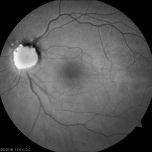 Fundus Autofluorescence of Optic Disc Drusen
Apr 26 2018 by Ahmad B. Tarabishy, MD
Fundus photographs and autofluorescence of a 75-year-old man with an epiretinal membrane in the left eye. Incidentally, he had a history of optic disc drusen, which show a striking hyperautofluorescence on FAF imaging.
Photographer: Michelle Howarth, Lakeland Eye Clinic
Imaging device: Zeiss Visucam
Condition/keywords: fundus autofluorescence (FAF), optic disc drusen
-
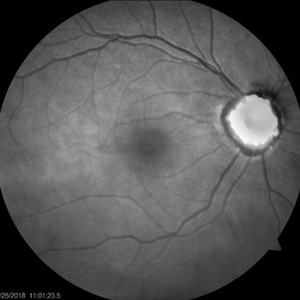 Fundus Autofluorescence of Optic Disc Drusen
Apr 26 2018 by Ahmad B. Tarabishy, MD
Fundus photographs and autofluorescence of a 75-year-old man with an epiretinal membrane in the left eye. Incidentally, he had a history of optic disc drusen, which show a striking hyperautofluorescence on FAF imaging.
Photographer: Michelle Howarth, Lakeland Eye Clinic
Imaging device: Zeiss Visucam
Condition/keywords: fundus autofluorescence (FAF), optic disc drusen
-
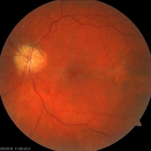 Color Fundus Photographs of Optic Disc Drusen
Apr 26 2018 by Ahmad B. Tarabishy, MD
Fundus photographs and autofluorescence of a 75-year-old man with an epiretinal membrane in the left eye. Incidentally, he had a history of optic disc drusen, which show a striking hyperautofluorescence on FAF imaging.
Photographer: Michelle Howarth, Lakeland Eye Clinic
Imaging device: Zeiss Visucam
Condition/keywords: fundus autofluorescence (FAF), optic disc drusen
A project from the American Society of Retina Specialists