-
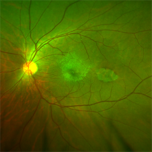
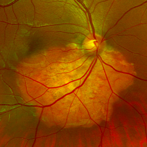
 Class 1A Choroidal Melanoma
Mar 10 2020 by David L Kilpatrick, MD
Fundus photograph of a 28-year-old female with a choroidal melanoma in the posterior pole. The presenting vision was 20/25. Transvitreal biopsy revealed a class 1A / PRAME negative tumor (Castle Biosciences). Based on the genetic expression profile, she will undergo TTT as the initial treatment modality.
Photographer: Retina Consultants of Alabama, P. C.
Condition/keywords: class 1A
-
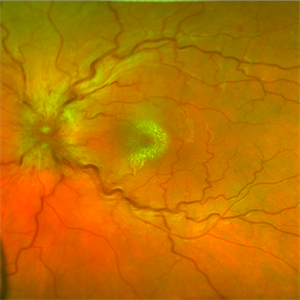
 Choroidal Osteoma
Mar 10 2020 by David L Kilpatrick, MD
Fundus photograph demonstrating a choroidal osteoma in the posterior pole. This patient had a drop in vision from 20/30 to 20/60 secondary to a new CNV that was treated with intravitreal bevacizumab.
Photographer: Retina Consultants of Alabama, P. C.
Condition/keywords: choroidal osteoma
-
 Torpedo Maculopathy
Apr 23 2020 by David L Kilpatrick, MD
50-year-old male with an asymptomatic ERM and adjacent torpedo maculopathy.
Photographer: Retina Consultants of Alabama
Imaging device: Optos Fundus Camera
Condition/keywords: epiretinal membrane (ERM), torpedo maculopathy
-
 Idiopathic Neuroretinitis
May 6 2020 by David L Kilpatrick, MD
Fundus photo of a 25-year-old white female with idiopathic neuroretinitis.
Photographer: RCA
Imaging device: Optos
Condition/keywords: neuroretinitis
-
 Retinal Cavernous Hemangioma
Nov 6 2020 by David L Kilpatrick, MD
15-year-old female with an asymptomatic retinal cavernous hemangioma.
Photographer: MS Retina Associoates
Imaging device: Optos
Condition/keywords: cavernous hemangioma of the retina
-
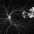
 CRAO with Cilioretinal Artery Sparing
Feb 11 2021 by David L Kilpatrick, MD
Patient presented with acute vision loss in the left eye. Exam showed CRAO with cilioretinal artery sparing. FA demonstrated retrograde venous filling.
Photographer: MS Retina Associoates
Imaging device: Optos
Condition/keywords: central retinal artery occlusion (CRAO), cilioretinal sparing
-
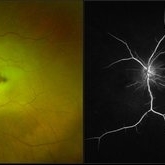
 Toxoplasmic Retinitis
Feb 11 2021 by David L Kilpatrick, MD
A 50-year-old female presented with a history of subacute central vision loss involving the right eye. Exam showed a focal, creamy white retinitis involving the fovea with mild vitritis. FA demonstrates blockage of the lesion initially followed by progressive leakage, most prominently at the border of the retinitis. Toxoplasma IgM and IgG were both markedly elevated. The patient was treated with a combination of Bactrim, Intravitreal Clindamycin, and Oral steroids.
Photographer: MS Retina Associoates
Imaging device: Optos
Condition/keywords: toxoplasmosis chorioretinitis, toxoplasmosis retinitis
-
 Choroidal Osteoma
Feb 23 2021 by David L Kilpatrick, MD
24-year-old female with an asymptomatic choroidal osteoma.
Photographer: MS Retina Associoates / Kyle McClellan
Imaging device: Optos
Condition/keywords: choroidal osteoma
-
 CRVO
CRVO
Apr 15 2021 by David L Kilpatrick, MD
A CRVO in a 72-year-old female with a history of hypertension.
Photographer: Kyle McClellan
Imaging device: Optos
Condition/keywords: central retinal vein occlusion (CRVO)
A project from the American Society of Retina Specialists