-
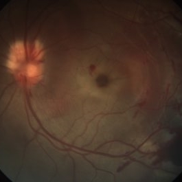 Traumatic Macular Hole
May 8 2021 by Jazli Tan
Fundus photograph of a 31-year-old man with a traumatic macular hole from a badminton racket injury. Fundoscopic examination revealed macular hole with associated sub-retinal, sub-retinal pigment epithelium, and sub-hyaloid bleed temporal to macula. Myelinated nerve fibre layer of the optic disc was noted superiorly with splinter hemorrhages.
Condition/keywords: traumatic macular hole
-
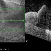 Optical Coherence Tomography
May 8 2021 by Jazli Tan
Optical Coherence Tomography (OCT) of a 31-year-old man with a traumatic macular hole from a badminton racket injury. OCT with enhanced depth imaging showed full thickness macular hole with subretinal fluid.
Condition/keywords: traumatic macular hole
-
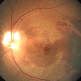 Fundus Photograph (Follow-up)
May 8 2021 by Jazli Tan
Follow-up fundoscopic examination revealed decreased subretinal blood and oedema at one week post injury, with persistent macular hole.
Condition/keywords: traumatic macular hole
A project from the American Society of Retina Specialists