-
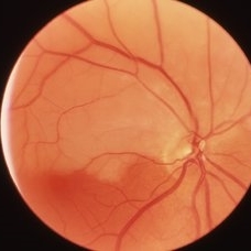
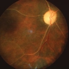 Old Central Retinal Artery Occlusion
Mar 26 2019 by Gary R. Cook, MD, FACS
Vascular and disc changes of a remote central retinal artery occlusion OD.
Condition/keywords: central retinal artery occlusion (CRAO)
-
 Embolic Central Retinal Artery Occlusion
Mar 26 2019 by Gary R. Cook, MD, FACS
58-year-old WM with embolic CRAO demonstrating a a cherry-red spot in macula, retinal whitening around the fovea, and the embolus in a inferotemporal branch retinal arteriole; VA= HM 6''
Imaging device: Topcon VT-50
Condition/keywords: central retinal artery occlusion (CRAO), cherry red spot, embolus, retinal whitening
-
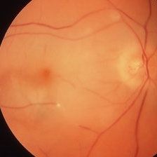
 Central Retinal Artery Occlusion
Mar 26 2019 by Gary R. Cook, MD, FACS
61-year-old male patient with acute CRAO OS demonstrating a hyperemic optic disc with a couple of peripapillary hemorrhages, generalized arteriolar narrowing, a cherry-red spot in the macula, and retinal whitening surrounding the fovea; VA= LP.
Imaging device: Topcon VT-50
Condition/keywords: central retinal artery occlusion (CRAO), cherry red spot, retinal whitening
-
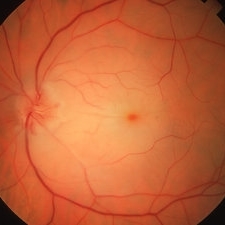
 Central Retinal Artery Occlusion with Cilioretinal Sparing
Mar 26 2019 by Gary R. Cook, MD, FACS
81-year-old Vietnamese female with acute CRAO with cilioretinal artery sparing OS; VA= 20/60.
Imaging device: Topcon VT-50
Condition/keywords: central retinal artery occlusion (CRAO), cilioretinal sparing, retinal whitening
-
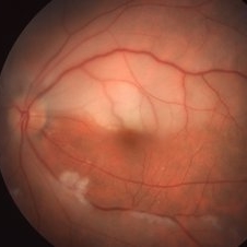
 Hemi-CRAO
Mar 26 2019 by Gary R. Cook, MD, FACS
Right eye of a white female showing an embolic superior hemi-central retinal artery occlusion with the embolus visible at the main artery bifurcation on the optic disc.
Condition/keywords: central retinal artery occlusion (CRAO), embolus
-
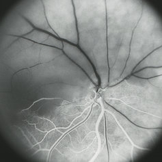 Hemi-CRAO
Mar 26 2019 by Gary R. Cook, MD, FACS
Mid-phase (laminar venous return) fluorescein angiogram image of an embolic superior hemi-CRAO showing marked delay in filling of the superior retinal arteriolar and venous vasculature and total loss of the retinal capillary bed in the superior hemisphere OD.
Condition/keywords: capillary closure, capillary nonperfusion, central retinal artery occlusion (CRAO), FA mid phase, fluorescein angiogram (FA)
A project from the American Society of Retina Specialists