-
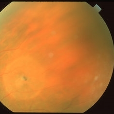
 Retinal Hole
Mar 26 2019 by Gary R. Cook, MD, FACS
63-year-old patient with retinal (macular) hole s/p laser photocoagulation for CNVM OS; VA= counting fingers at 6 feet.
Imaging device: Topcon VT-50
Condition/keywords: argon photocoagulation, choroidal neovascular membrane (CNVM), complication, laser photocoagulation, retinal hole
-
 Operculated Retinal Hole
Apr 8 2019 by Gary R. Cook, MD, FACS
White female with an operculated retinal hole with a small cuff of surrounding SRF; V.A. = 20/25
Imaging device: Topcon VT-50
Condition/keywords: operculated retinal hole
-
 Retinal Hole with Subclinical Detachment
Apr 8 2019 by Gary R. Cook, MD, FACS
64-year-old white female with an asymptomatic retinal hole with some pigment and a 1DD surrounding cuff of subretinal fluid; V.A. = 20/20-3.
Imaging device: Topcon VT-50
Condition/keywords: retinal hole, subclinical detachment
-
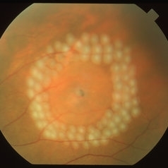 Retinal Hole with Subclinical Detachment
Apr 8 2019 by Gary R. Cook, MD, FACS
Immediate post laser photocoagulation treatment of a retinal hole with a 1DD surrounding subclinical detachment in a 64-year-old white female; V.A. = 20/20-3.
Imaging device: Topcon VT-50
Condition/keywords: laser photocoagulation, laser retinopexy, post-laser, retinal hole, subclinical detachment
A project from the American Society of Retina Specialists