-
 Regional Choriocapillaris Atrophy
Jun 18 2019 by Gary R. Cook, MD, FACS
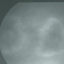
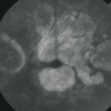
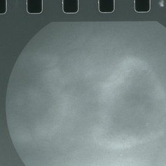
Late-phase (5 minutes) FA frame of the left eye of a 73-year-old white female with regional choriocapillaris atrophy showing light staining from intact choriocapillaris around the margins of the peripapillary and macular areas of RPE and choriocapillaris atrophy; V.A. = 20/100
Imaging device: Topcon VT-50
Condition/keywords: atrophy, choriocapillaris, FA late phase, hereditary choroidal atrophy, hereditary choroidal dystrophy
-
 Central Areolar Choriocapillaris Atrophy
Mar 26 2019 by Gary R. Cook, MD, FACS
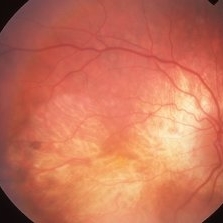
Right eye of a 64-year-old male with central (or regional) atrophy of the RPE and choriocapillaris in the macula; VA= 20/30.
Imaging device: Topcon VT-50
Condition/keywords: choriocapillaris, hereditary choroidal atrophy, hereditary choroidal dystrophy
-
 Central Areolar Choriocapillaris Atrophy
Mar 26 2019 by Gary R. Cook, MD, FACS
Late-phase fluorescein angiogram image of the right eye of a 64-year-old white male with central areolar choriocapillaris atrophy showing late leakage from intact choriocapillaris around the perimeter of the disc and macular areas of choriocapillaris atrophy; VA= 20/50
Imaging device: Topcon VT-50
Condition/keywords: FA late phase, hereditary choroidal atrophy, hereditary choroidal dystrophy
-
 Central Areolar Choriocapillaris Atrophy
Mar 26 2019 by Gary R. Cook, MD, FACS
Left eye of a 64-year-old male with central (regional) areolar choroidal dystrophy showing fairly well circumscribed atrophy of the RPE and choriocapillaris in the macula; VA= 20/30
Imaging device: Topcon VT-50
Condition/keywords: choriocapillaris, hereditary choroidal atrophy, hereditary choroidal dystrophy
-
 Central Areolar Choriocapillaris Atrophy
Mar 26 2019 by Gary R. Cook, MD, FACS
Late-phase fluorescein angiogram image of the left eye of a 64-year-old white male with central areolar choriocapillaris atrophy showing light late staining of the central lesions OS; V.A. = 20/30
Imaging device: Topcon VT-50
Condition/keywords: choriocapillaris, FA late phase, hereditary choroidal atrophy, hereditary choroidal dystrophy
-
 Regional Choriocapillaris Atrophy
Mar 26 2019 by Gary R. Cook, MD, FACS
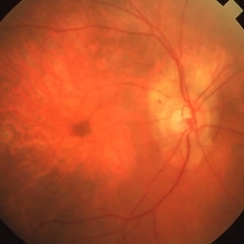
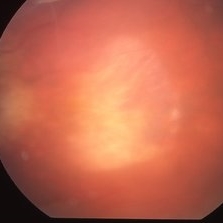
73-year-old white female with regional choriocapillaris atrophy in the posterior pole of her right eye; VA= 20/100.
Imaging device: Topcon VT-50
Condition/keywords: atrophy, choriocapillaris, hereditary choroidal atrophy, hereditary choroidal dystrophy
-
 Regional Choriocapillaris Atrophy
Mar 26 2019 by Gary R. Cook, MD, FACS
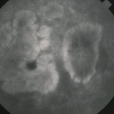
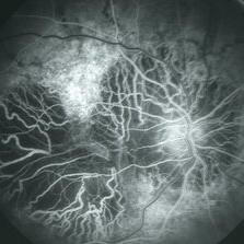
Mid-phase (laminar venous return phase) FA frame demonstrating thinning/loss of the RPE, choriocapillaris loss, and increased visibility of the larger choroidal vessels around the disc and in the macula from a 73-year-old white female with regional choriocapillaris atrophy; VA= 20/100.
Imaging device: Topcon VT-50
Condition/keywords: atrophy, choriocapillaris, hereditary choroidal atrophy, hereditary choroidal dystrophy
-
 Regional Choriocapillaris Atrophy
Mar 26 2019 by Gary R. Cook, MD, FACS
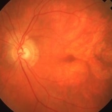
73-year-old white female with regional choriocapillaris atrophy OS; VA= 20/100.
Imaging device: Topcon VT-50
Condition/keywords: atrophy, choriocapillaris, hereditary choroidal atrophy, hereditary choroidal dystrophy
-
 Regional Choriocapillaris Atrophy
Mar 26 2019 by Gary R. Cook, MD, FACS
Late-phase (5 minutes) FA frame of the left eye of a 73-year-old white female with regional choriocapillaris atrophy showing light staining from intact choriocapillaris around the margins of the peripapillary and macular areas of RPE and choriocapillaris atrophy; VA= 20/100.
Imaging device: Topcon VT-50
Condition/keywords: atrophy, choriocapillaris, FA late phase, hereditary choroidal atrophy, hereditary choroidal dystrophy
A project from the American Society of Retina Specialists