-
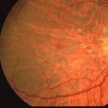 Bilateral C-R Folds
Mar 26 2019 by Gary R. Cook, MD, FACS
Fundus photo of the right eye of a white male with bilateral C-R folds.
Imaging device: Topcon VT-50
Condition/keywords: bilateral chorioretinal folds, chorioretinal fold
-
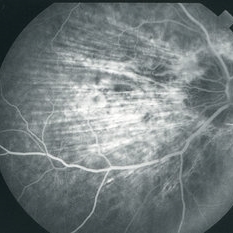 C-R Folds
Mar 26 2019 by Gary R. Cook, MD, FACS
Mid-phase FA image of the right eye of a white male with bilateral C-R folds showing alternating hyper- and hypofluorescent bands.
Imaging device: Topcon VT-50
Condition/keywords: bilateral chorioretinal folds, chorioretinal fold, FA mid phase, fluorescein angiogram (FA)
-
 Bilateral C-R Folds
Mar 26 2019 by Gary R. Cook, MD, FACS
Fundus photo of the left eye of a white male with bilateral C-R folds.
Imaging device: Topcon VT-50
Condition/keywords: bilateral chorioretinal folds, chorioretinal fold
-
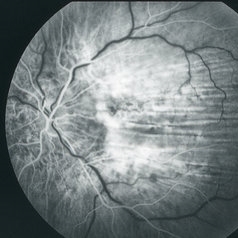 C-R Folds
Mar 26 2019 by Gary R. Cook, MD, FACS
Early phase FA frame of the left eye of a WM with bilateral C-R folds showing alternating hyper- and hypofluorescent bands.
Imaging device: Topcon VT-50
Condition/keywords: bilateral chorioretinal folds, chorioretinal fold, FA early phase, fluorescein angiogram (FA)
A project from the American Society of Retina Specialists