-
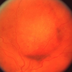
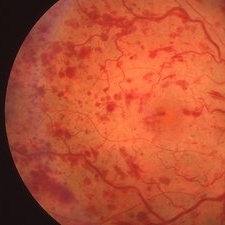 Central Retinal Vein Occlusion
Mar 26 2019 by Gary R. Cook, MD, FACS
56-year-old white female with CRVO (intermediate perfusion status) with macular edema OD showing dilated and tortuous venous vasculature, and dot, blot and NFL hemorrhages in all 4 quadrants; VA=20/400.
Imaging device: Topcon VT-50
Condition/keywords: central retinal vein occlusion (CRVO)
-
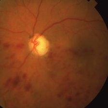
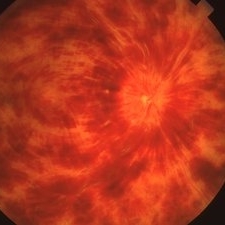 Progression of CRVO
Mar 26 2019 by Gary R. Cook, MD, FACS
The same 56-year-old white female 6 weeks later showing marked progression to non-perfused status with a hyperemic optic disc and marked and extensive retinal hemorrhages: VA= 20/2000.
Imaging device: Topcon VT-50
Condition/keywords: central retinal vein occlusion (CRVO)
-
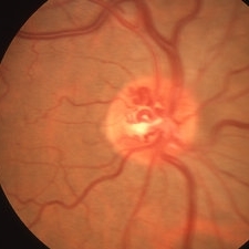 Disc Collaterals in CRVO
Mar 27 2019 by Gary R. Cook, MD, FACS
Elderly white male with remote, compensated CRVO showing persistent dilation of the venous vasculature and optic disc collaterals; V.A.= 20/20
Imaging device: Topcon VT-50
Condition/keywords: central retinal vein occlusion (CRVO), collaterals, disc
-
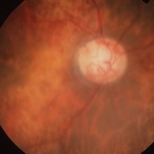 CRVO with Disc Collaterals
Mar 27 2019 by Gary R. Cook, MD, FACS
81-year-old white female with remote CRVO with disc collaterals OD; V.A.= counting fingers 1 ft.
Imaging device: Topcon VT-50
Condition/keywords: central retinal vein occlusion (CRVO), collaterals, disc
-
 Retinal Cyst after CRVO
Mar 27 2019 by Gary R. Cook, MD, FACS
Elderly white female s/p CRVO 3 years ago now with retinal cyst OD; V.A.= hand motions.
Condition/keywords: central retinal vein occlusion (CRVO), retinal cyst
-
 Hemi-CRVO
Mar 27 2019 by Gary R. Cook, MD, FACS
78-year-old African American female patient with COAG and ischemic inferior hemi-CRVO OS; V.A. = HM 1 ft.
Imaging device: Topcon VT-50
Condition/keywords: chronic open-angle glaucoma (COAG), hemi CRVO, retinal hemorrhage
-
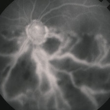 Hemi-CRVO
Jun 4 2019 by Gary R. Cook, MD, FACS
Mid-phase FA image of 78-year-old African American female patient with COAG and a very ischemic inferior hemi-CRVO showing loss of almost all of the capillary bed and staining of the veins in the lower hemisphere of the left eye; V.A. = HM 1 ft.
Imaging device: Topcon VT-50
Condition/keywords: central retinal vein occlusion (CRVO), FA mid phase, fluorescein angiogram (FA), hemi CRVO, ischemia, ischemic CRVO
A project from the American Society of Retina Specialists