-
 Slide 14-3
Mar 4 2019 by Lancaster Course in Ophthalmology
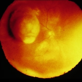
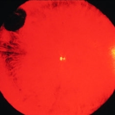
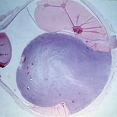
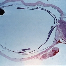
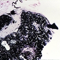
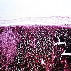
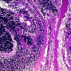
Among the objective manifestations of a choroidal melanoma, the most important is the presence of a solid mass seen on ophthaloscopic presentation. (seen in Slides 14-1 to 14-13). The shape and projection of the tumor can be best appreciated by observation with the indirect ophthalmoscope. The appearance of these tumors is typically that of a mushroom-shaped, collar button-shaped mass.
Condition/keywords: melanoma, tumor
-
 Slide 14-4
Mar 4 2019 by Lancaster Course in Ophthalmology
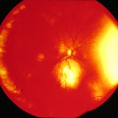
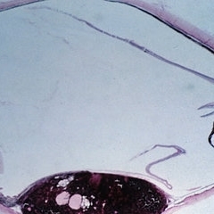
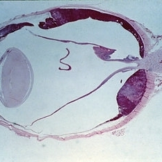
Among the objective manifestations of a choroidal melanoma, the most important is the presence of a solid mass seen on ophthaloscopic presentation. (seen in Slides 14-1 to 14-13). The shape and projection of the tumor can be best appreciated by observation with the indirect ophthalmoscope. The appearance of these tumors is typically that of a mushroom-shaped, collar button-shaped mass.
Condition/keywords: melanoma, tumor
-
 Slide 14-5
Mar 4 2019 by Lancaster Course in Ophthalmology
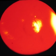
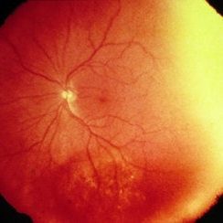
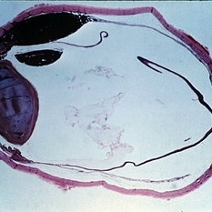
Among the objective manifestations of a choroidal melanoma, the most important is the presence of a solid mass seen on ophthaloscopic presentation. (seen in Slides 14-1 to 14-13). The shape and projection of the tumor can be best appreciated by observation with the indirect ophthalmoscope. The appearance of these tumors is typically that of a mushroom-shaped, collar button-shaped mass.
Condition/keywords: melanoma, tumor
-
 Slide 14-6
Mar 4 2019 by Lancaster Course in Ophthalmology
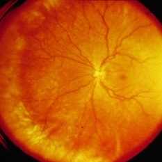
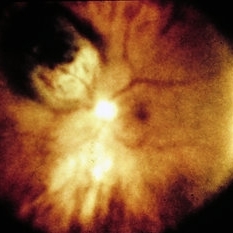
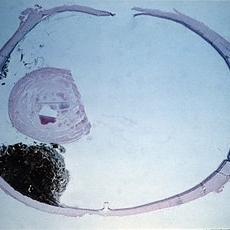
Among the objective manifestations of a choroidal melanoma, the most important is the presence of a solid mass seen on ophthaloscopic presentation. (seen in Slides 14-1 to 14-13). The shape and projection of the tumor can be best appreciated by observation with the indirect ophthalmoscope. The appearance of these tumors is typically that of a mushroom-shaped, collar button-shaped mass.
Condition/keywords: melanoma, tumor
-
 Slide 14-7
Mar 4 2019 by Lancaster Course in Ophthalmology
Among the objective manifestations of a choroidal melanoma, the most important is the presence of a solid mass seen on ophthaloscopic presentation. (seen in Slides 14-1 to 14-13). The shape and projection of the tumor can be best appreciated by observation with the indirect ophthalmoscope. The appearance of these tumors is typically that of a mushroom-shaped, collar button-shaped mass.
Condition/keywords: melanoma, tumor
-
 Slide 14-8
Mar 14 2019 by Lancaster Course in Ophthalmology
Among the objective manifestations of a choroidal melanoma, the most important is the presence of a solid mass seen on ophthaloscopic presentation. (seen in Slides 14-1 to 14-13). The shape and projection of the tumor can be best appreciated by observation with the indirect ophthalmoscope. The appearance of these tumors is typically that of a mushroom-shaped, collar button-shaped mass.
Condition/keywords: melanoma, tumor
-
 Slide 14-2
Mar 4 2019 by Lancaster Course in Ophthalmology
Among the objective manifestations of a choroidal melanoma, the most important is the presence of a solid mass seen on ophthaloscopic presentation. (seen in Slides 14-1 to 14-13). The shape and projection of the tumor can be best appreciated by observation with the indirect ophthalmoscope. The appearance of these tumors is typically that of a mushroom-shaped, collar button-shaped mass.
Condition/keywords: melanoma, tumor
-
 Slide 14-1
Mar 4 2019 by Lancaster Course in Ophthalmology
Among the objective manifestations of a choroidal melanoma, the most important is the presence of a solid mass seen on ophthaloscopic presentation. (seen in Slides 14-1 to 14-13). The shape and projection of the tumor can be best appreciated by observation with the indirect ophthalmoscope. The appearance of these tumors is typically that of a mushroom-shaped, collar button-shaped mass.
Condition/keywords: melanoma, tumor
-
 Slide 14-9
Mar 4 2019 by Lancaster Course in Ophthalmology
Among the objective manifestations of a choroidal melanoma, the most important is the presence of a solid mass seen on ophthaloscopic presentation. (seen in Slides 14-1 to 14-13). The shape and projection of the tumor can be best appreciated by observation with the indirect ophthalmoscope. The appearance of these tumors is typically that of a mushroom-shaped, collar button-shaped mass.
Condition/keywords: melanoma, tumor
-
 Slide 14-10
Mar 4 2019 by Lancaster Course in Ophthalmology
Among the objective manifestations of a choroidal melanoma, the most important is the presence of a solid mass seen on ophthaloscopic presentation. (seen in Slides 14-1 to 14-13). The shape and projection of the tumor can be best appreciated by observation with the indirect ophthalmoscope. The appearance of these tumors is typically that of a mushroom-shaped, collar button-shaped mass.
Condition/keywords: melanoma, tumor
-
 Slide 14-11
Mar 4 2019 by Lancaster Course in Ophthalmology
Among the objective manifestations of a choroidal melanoma, the most important is the presence of a solid mass seen on ophthaloscopic presentation. (seen in Slides 14-1 to 14-13). The shape and projection of the tumor can be best appreciated by observation with the indirect ophthalmoscope. The appearance of these tumors is typically that of a mushroom-shaped, collar button-shaped mass.
Condition/keywords: melanoma, tumor
-
 Slide 14-12
Mar 4 2019 by Lancaster Course in Ophthalmology
Among the objective manifestations of a choroidal melanoma, the most important is the presence of a solid mass seen on ophthaloscopic presentation. (seen in Slides 14-1 to 14-13). The shape and projection of the tumor can be best appreciated by observation with the indirect ophthalmoscope. The appearance of these tumors is typically that of a mushroom-shaped, collar button-shaped mass.
Condition/keywords: melanoma, tumor
-
 Slide 14-13
Mar 4 2019 by Lancaster Course in Ophthalmology
Among the objective manifestations of a choroidal melanoma, the most important is the presence of a solid mass seen on ophthaloscopic presentation. (seen in Slides 14-1 to 14-13). The shape and projection of the tumor can be best appreciated by observation with the indirect ophthalmoscope. The appearance of these tumors is typically that of a mushroom-shaped, collar button-shaped mass.
Condition/keywords: melanoma, tumor
-
 Slide 14-14
Mar 4 2019 by Lancaster Course in Ophthalmology
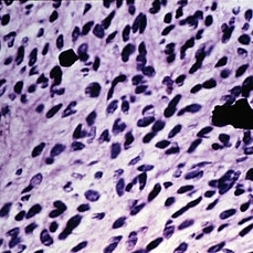
Spindle A melanomas are composed of cohesive cells with small, spindle-shaped nuclei containing a dark stripe contributed by a nuclear fold. The nuclei are not distinct. The cytoplasm is ill-defined, and the cell borders are not easily identified. They comprise approximately 5 percent of ciliary body and choroidal melanomas.
Condition/keywords: melanoma, spindle A melanoma
-
 Slide 14-15
Mar 4 2019 by Lancaster Course in Ophthalmology
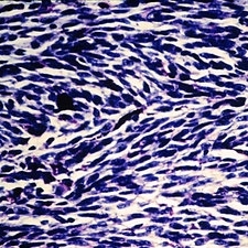
Spindle B and fascicular melanomas are made up of cohesive cells having distinct spindle-shaped nuclei with prominent nucleoli. The cytoplasm is indistinct, and the cell borders are not clearly seen by light microscopy. This cell type comprises 39 percent of ciliary body and choroidal melanomas. The fascicular type is a subgroup of spindle B melanomas in which the spindle-shaped cells form a pallisaded arrangement termed a fascicular pattern. They account for approximately 6 percent of choroidal melanomas.
Condition/keywords: melanoma, spindle B melanoma
-
 Slide 14-16
Mar 4 2019 by Lancaster Course in Ophthalmology
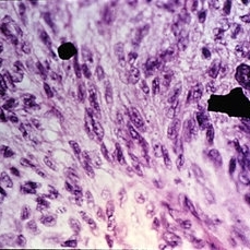
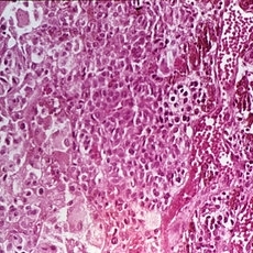
Epithelioid melanomas are composed of noncohesive, large cells and round nuclei and prominent nucleoli; abundant eosinophilic cytoplasm and well-demarcated cell borders are present. This is the rarest type (3 percent) of ciliary and choroidal melanomas.
Condition/keywords: cytoplasm, eosinophilic, melanoma
-
 Slide 14-17
Mar 4 2019 by Lancaster Course in Ophthalmology
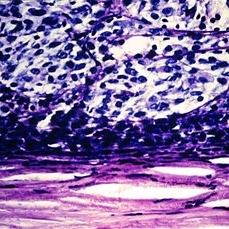
Mixed cell type consists of a neoplasm showing a significant spindle cell population (usually spindle B) together with numerous epithelioid components. It should be noted that the term 'mixed cell type does not refer to tumors composed of spindle A and spindle B cells but rather tumors containing spindle cell and epithelioid cells. This is the most common type of uveal and choroidal melanoma, accounting for 45 percent of lesions.
Condition/keywords: melanoma
-
 Slide 14-18
Mar 4 2019 by Lancaster Course in Ophthalmology
The most convincing evidence for the relationship of uveal nevi to malignant melanoma is the histologic observation of presumed nevus cells in association with ocular malignant melanomas in enucleated eyes.
Condition/keywords: malignant melanoma, melanoma
-
 Slide 14-19
Mar 4 2019 by Lancaster Course in Ophthalmology
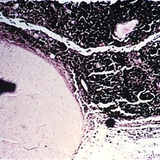
Ciliary body melanomas may appear early as a discrete mass or may show a diffuse growth pattern. As they enlarge, they may grow anteriorly onto the iris or extend posteriorly to the choroid.
Condition/keywords: ciliary body melanoma, melanoma
-
 Slide 14-20
Mar 4 2019 by Lancaster Course in Ophthalmology
Ciliary body melanomas may appear early as a discrete mass or may show a diffuse growth pattern. As they enlarge, they may grow anteriorly onto the iris or extend posteriorly to the choroid.
Condition/keywords: ciliary body melanoma, melanoma
-
 Slide 14-21
Mar 4 2019 by Lancaster Course in Ophthalmology
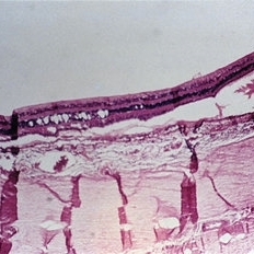
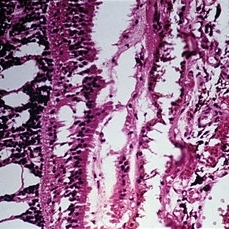
Cystoid degeneration of the overlying retina has been noted in early most choroidal melanomas.
Condition/keywords: cystoid macular degeneration, melanoma
-
 Slide 14-22
Mar 4 2019 by Lancaster Course in Ophthalmology
Cystoid degeneration of the overlying retina has been noted in early most choroidal melanomas.
Condition/keywords: cystoid macular degeneration, melanoma
-
 Slide 14-23
Mar 4 2019 by Lancaster Course in Ophthalmology
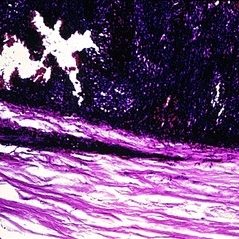
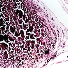
Choroidal melanomas, particularly in the macular region, invaginate or become "embedded" in the underlying sclera. This results in thinning and slight ectasia of the sclera but in most case no actual tumor invasion. This tendency has clinical significance in that the tumor may obtain an appreciable size but have minimal elevation, and its dimensions may not be appreciated ophthalmoscopically. Direct invasion of the sclera occurs in about 15 percent of tumors.
Condition/keywords: melanoma, sclera, scleral indentation
-
 Slide 14-24
Mar 4 2019 by Lancaster Course in Ophthalmology
In advanced cases, melanomas may present as an orbital or subconjunctival mass.
Condition/keywords: melanoma
-
 Slide 14-25
Mar 4 2019 by Lancaster Course in Ophthalmology
In advanced cases, melanomas may present as an orbital or subconjunctival mass.
Condition/keywords: melanoma
-
 Slide 14-26
Mar 4 2019 by Lancaster Course in Ophthalmology
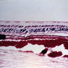
Other lesions mistaken for melanomas such as serous detachments of the retina and choroid.
Condition/keywords: melanoma, serous detachment
-
 Slide 14-27
Mar 4 2019 by Lancaster Course in Ophthalmology
Other lesions mistaken for melanomas such as serous detachments of the retina and choroid.
Condition/keywords: melanoma, serous detachment
-
 Slide 14-28
Mar 4 2019 by Lancaster Course in Ophthalmology
Other lesions mistaken for melanomas such as other tumors including hemangiomas, metastic tumors, melanocytoma of the disc, nevus of the choroid, hypertrophy of the pigment epithelium, adenoma and adenocarcinoma of the pigment epithelium, reactive proliferation of the pigment epithelium, and lymphoma and leukemia.
Condition/keywords: choroidal nevus, hemangioma, leukemia, lymphoma, melanoma, optic disc melanocytoma, retinal pigment epithelium (RPE) hypertrophy
-
 Slide 14-29
Mar 4 2019 by Lancaster Course in Ophthalmology
Other lesions mistaken for melanomas such as hemorrhages which may be choroidal, subpigment epithelial, subretinal, intraretinal, or vitreous.
Condition/keywords: hemorrhage, melanoma
-
 Slide 14-30
Mar 4 2019 by Lancaster Course in Ophthalmology
The vast majority of iris melanomas are composed of spindle A and/or spindle B cells and are relatively benign.
Condition/keywords: iris melanoma, melanoma, spindle A melanoma, spindle B melanoma, spindle cells
-
 Slide 14-31
Mar 4 2019 by Lancaster Course in Ophthalmology
The vast majority of iris melanomas are composed of spindle A and/or spindle B cells and are relatively benign.
Condition/keywords: iris melanoma, melanoma, spindle A melanoma, spindle B melanoma, spindle cells
A project from the American Society of Retina Specialists