-
 Retinal Crystals in Macular Telangiectasia
Jan 6 2019 by John S. King, MD
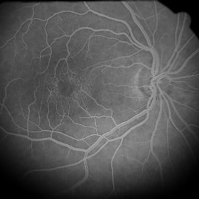
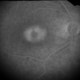
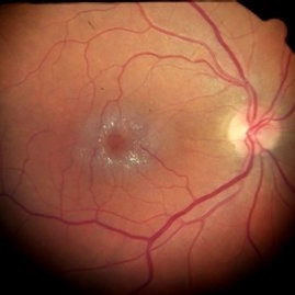
61-year-old white female, healthy, slow progressive central vision loss OD. 20/200 OD and 20/30 OS with mild-mod NSC OU. Retinal crystals around fovea in the posterior pole. FA shows 360 degrees of parafoveal telangiectatic vessels that leak without signs of CNVM. Minimal changes in fellow eye. Asymmetric MacTel 2. Monitoring and using AREDS 2.
Photographer: Kay Dalby
Imaging device: Topcon 50 (satellite office)
Condition/keywords: crystalline retinopathy, cystoid macular edema (CME), macular telangiectasia
-
 Retinal Crystals in Macular Telangiectasia
Jan 6 2019 by John S. King, MD
61-year-old white female, healthy, slow progressive central vision loss OD. 20/200 OD and 20/30 OS with mild-mod NSC OU. Retinal crystals around fovea in the posterior pole. FA shows 360 degrees of parafoveal telangiectatic vessels that leak without signs of CNVM. Minimal changes in fellow eye. Asymmetric MacTel 2. Monitoring and using AREDS 2.
Photographer: Kay Dalby
Imaging device: Topcon 50 (satellite office)
Condition/keywords: crystalline retinopathy, cystoid macular edema (CME), macular telangiectasia
A project from the American Society of Retina Specialists