-
 BDUMP
BDUMP
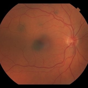
Dec 11 2018 by John S. King, MD
67-year-old white female with normal vision four months ago, consulted for dry AMD. She reported that vision in the left eye had worsened over the last two months and had progressively gotten worse. Denied history of cancer, or her primary eye doctor ever mentioning choroidal nevi. Va cc was 20/30 OD and 20/100 OS. No RAPD. IOP 9-10 OU. Anterior segment had some stellate like pigmented dusting of the endothlium, a/c was quiet, 2+NSC OU. Vitreous quiet; multiple, flat, pigmented choroidal lesions varying in size was seen the in fundus. Area in the temporal macula extending up to the superior arcade in the left eye that was suspicious for a mass; it did have a "giraffe like" pattern on one of the early FA pics; the OCT in this area showed thickening of the choroid without a definite mass lesion, and overlying thickening of the RPE, or exudative like scar, with SRF directly above. Sent patient to Dr. Matt Wilson, who confirmed BDUMP, and had patient sent to oncology to find a possible primary lesion. Mass seen on CT chest; biopsy revealed non-small cell lung cancer, and is getting chemo/radio treatment. Ocular findings have not progressed over the last few months.
Photographer: Stacey Coleman
Imaging device: Topcon 50
Condition/keywords: bilateral diffuse uveal melanocytic proliferation (BDUMP)
-
 BDUMP
BDUMP
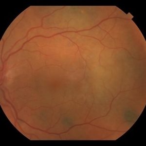
Dec 11 2018 by John S. King, MD
67-year-old white female with normal vision four months ago, consulted for dry AMD. She reported that vision in the left eye had worsened over the last two months and had progressively gotten worse. Denied history of cancer, or her primary eye doctor ever mentioning choroidal nevi. Va cc was 20/30 OD and 20/100 OS. No RAPD. IOP 9-10 OU. Anterior segment had some stellate like pigmented dusting of the endothlium, a/c was quiet, 2+NSC OU. Vitreous quiet; multiple, flat, pigmented choroidal lesions varying in size was seen the in fundus. Area in the temporal macula extending up to the superior arcade in the left eye that was suspicious for a mass; it did have a "giraffe like" pattern on one of the early FA pics; the OCT in this area showed thickening of the choroid without a definite mass lesion, and overlying thickening of the RPE, or exudative like scar, with SRF directly above. Consulted with Dr. Matt Wilson, who confirmed diagnosis, and had patient evaluated by oncology, who diagnosed non-small cell lung cancer.
Photographer: Stacey Coleman
Imaging device: Topcon
Condition/keywords: bilateral diffuse uveal melanocytic proliferation (BDUMP)
A project from the American Society of Retina Specialists