-
 Silicon Droplet in the Cup of Optic Nerve After Multiple Intravitreal Avastin Treatments
Nov 1 2018 by Tammy Mclaughlin
Fundus photograph of a 68-year-old man with silicon droplet in the cup of optic nerve after multiple intravitreal Avastin treatments.
Photographer: Tammy Mclaughlin, Carolina Retina Center, Sumter SC
Imaging device: Zeiss Visucam
Condition/keywords: optic nerve
-
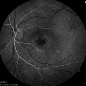
 Heart shaped Macular Edema
Nov 1 2018 by Tammy Mclaughlin
OCT of 65-year-old woman with macular edema.
Photographer: Tammy Mclaughlin, Carolina Retina Center, Sumter SC
Imaging device: Heidelberg
Condition/keywords: macular edema
-
 Multi-Color Image of Epiretinal Membrane With Prominent Edge Superior Temporal Macula
Nov 7 2018 by Tammy Mclaughlin
Multi-color image of female patient with epiretinal membrane OD. Clinical exam and diagnostic testing show large central ERM, retinal striae, and a prominent edge in the superior temporal macula. Visual acuity 20/25.
Photographer: Tammy Mclaughlin, Carolina Retina Center, Sumter SC 29150
Imaging device: Heidelberg
Condition/keywords: epiretinal membrane (ERM)
-
 Congenital Prepapillary Arterial Loop With a Figure-of-Eight Configuration
Mar 27 2019 by Tammy Mclaughlin
Congenital prepapillary arterial loop with a figure-of-eight configuration. OD. There is a twisted anomalous vessel eminating from the optic disc into the vitreous. Likely a congenital anomaly. Does not require treatment and should not be a vision threat.
Photographer: Tammy Mclaughlin, Carolina Retina Center 645 W. Wesmark Blvd. Sumter, Sc 29150
Condition/keywords: congenital anomaly, congenital prepapillary vascular loop
-
 Focal Retinitis- Unilateral Acute Idiopathic Maculopathy OS
Nov 13 2019 by Tammy Mclaughlin
Unilateral acute idiopathic maculopathy OS. No prodromal URI. Symptoms present for 1 1/2 weeks. No serous detachment presently. Does have typical circular RPE changes in macula without leakage or edema. Typical course is spontaneous improvement with residual RPE changes. Recommended observation.
Photographer: Tammy Mclaughlin
Imaging device: Zeiss Visucam
Condition/keywords: unilateral acute idiopathic maculopathy
-
 Focal Retinitis- Unilateral Acute Idiopathic Maculopathy OS.
Nov 13 2019 by Tammy Mclaughlin
No prodromal URI. Symptoms present for 1 1/2 weeks. No serous detachment presently. Does have typical circular RPE changes in macula without leakage or edema. Typical course is spontaneous improvement with residual RPE changes. Recommended observation.
Photographer: Tammy Mclaughlin
Imaging device: Zeiss Visucam
Condition/keywords: focal retinitis, unilateral acute idiopathic maculopathy
-
 Varices OS Prominent Vortex Vein Ampulla
Dec 18 2019 by Tammy Mclaughlin
There is a prominent vortex vein ampulla. Typically these are present in in the inferior nasal quadrant when the eye is directed toward the vessel. It is benign and not to be confused with a mass lesion. Reassured patient. Varices OU. Prominent Vortex Vein Ampulla. Recommended observation.
Photographer: Tammy Mclaughlin, Carolina Retina Center, Sumter SC
Condition/keywords: retinal varices, vortex vein
-
 Varices OD Prominent Vortex Vein Ampulla
Dec 18 2019 by Tammy Mclaughlin
There is a prominent vortex vein ampulla. Typically these are present in in the inferior nasal quadrant when the eye is directed toward the vessel. It is benign and not to be confused with a mass lesion. Reassured patient. Varices OU. Prominent Vortex Vein Ampulla. Recommended observation.
Photographer: Tammy Mclaughlin, Carolina Retina Center, Sumter SC
Condition/keywords: retinal varices, vortex vein
-
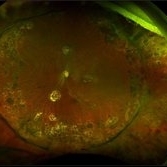 Scleral Buckle
Dec 31 2020 by Tammy Mclaughlin
Central serous chorioretinopathy OS (Incidental SRF 6/25/20 a little worse then stable). Repaired recurrent retinal detachment with PVR OS (new inferior starfold, anterior loop, and heavy pigment. s/p PPV 11/19/19, PPV/SBP/MP/SO 1/7/20). Posterior vitreous detachment OD. Floaters OD. Epiretinal membrane OS (mild).
Photographer: Tammy Mclaughlin, Carolina Retina Center, Sumter SC
Condition/keywords: scleral buckle
A project from the American Society of Retina Specialists