-
 Retina Dialysis Associated Retinal Detachment
Aug 22 2018 by Luis J Haddock, MD
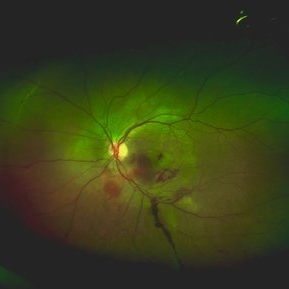
Optos color fundus photography showing nasal macula-on retinal detachment associated to a superonasal chronic retinal dialysis. This is a 19-year-old male who presented to glaucoma for elevated IOP. On dilated fundus exam retinal detachment was noted. Extended DFE showed nasal macula-on retinal detachment associated to a nasal retinal dialysis with peripheral vitreous contraction. The patient reported remote history of BB gun injury to his left eye at 5-years-old.
Imaging device: Optos California
Condition/keywords: blunt trauma, retinal dialysis
-
 Traumatic Macular Hemorrhage
Aug 22 2018 by Luis J Haddock, MD
16-year-old male with injury to left eye after a tennis ball was fired from a potato gun. The optos fundus photograph shows multilayered traumatic hemorrhage (preretinal, intraretinal and subretinal), there is choroidal rupture under the subretinal hemorrhage. The macula also shows contusion injury with Berlins edema. There is preretinal hemorrhage over the fovea and a macular hole cannot be ruled out.
Imaging device: Optos California
Condition/keywords: choroidal rupture, macular hemorrhage, trauma
-
 Traumatic Giant Retinal Tear Associated Retinal Detachment
Nov 9 2019 by Luis J Haddock, MD
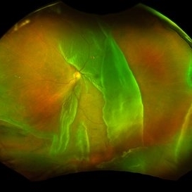
This wide field fundus photograph of the left eye shows a traumatic giant retinal tear associated with total retinal detachment. The image shows the torn superior retina folded over the macula with the underside of the retina visible. There is associated peripheral choroidal detachment due to hypotony from giant retinal tear. This patient has history of spondyloepithelial dysplasia with dwarfism and presented with vision loss after a recent blunt trauma with elbow to the eye.
Imaging device: Optos
Condition/keywords: giant retinal tear, traumatic optic neuropathy
-
 GRT Detachment of 10+ Clock Hours With Folded Retina
Sep 11 2025 by Luis J Haddock, MD
Fundus photo of giant retinal tear detachment involving 10+ hours of continuous tearing of the retina, visible anterior edge of retina over GRT.
Photographer: Natella Romero, University of Miami, Bascom Palmer Eye Institute
Imaging device: Optos
Condition/keywords: acute retinal detachment, Giant retinal tear
A project from the American Society of Retina Specialists