-
 Sea-Saw Vitreoretinal Dance
Mar 12 2021 by RUSHIK PATEL
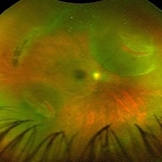
Fundus photograph of 50-year-old female showing posterior vitreous detachment, 2 retinal tears with localized retinal detachment exactly 180 degrees apart with optic disc in between as a Falcrum giving an appearance of sea saw retinal tears. Macula was attached with lattice retinal degeneration superiorly.
Photographer: Rushik Patel, Netralaya Super Speciality Eye Hospital
Condition/keywords: peripheral lattice degeneration, retinal tear
-
 Sub-ILM Hemorrhage (Dehemoglobinized + Red) - Valsalva Retinopathy
Jun 12 2021 by RUSHIK PATEL
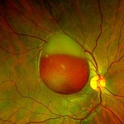
Fundus Image of 21-year-old boy with sub-ILM hemorrhage (dehemoglobinized + red ) following valsalva maneuver 10 days back.
Photographer: Rushik Patel, Netralaya Super Speciality Eye Hospital, Ahmedabad, Gujarat
Imaging device: Optos
Condition/keywords: sub-inner limiting membrane hemorrhage, valsalva retinopathy
-
 Rhegmatogenous Retinal Detachment
Jul 2 2021 by RUSHIK PATEL
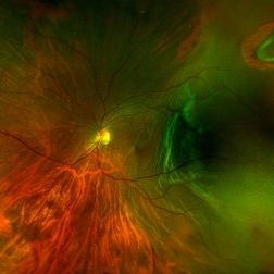
Fundus photograph of an 50-year-old female with rhegmatogenous retinal detachment, horseshoe tear.
Photographer: Rushik Patel, Netralaya Super Speciality Eye Hospital, Ahmedabad, Gujarat
-
 Retinal Holes, Demarcation Line
Jul 19 2021 by RUSHIK PATEL
Utlrawide pseudo-color fundus photograph of 28-year-old boy with 2 retinal hole surrounded by subretinal fluid less than 1 disc diameter and demarcation line.
Photographer: Rushik Patel, Netralaya Super Speciality Eye Hospital
Condition/keywords: retinal hole
-
 Reverse Polarity OCT Angiography of Proliferative Diabetic Retinopathy
Aug 31 2021 by RUSHIK PATEL
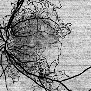
Reverse polarity OCTA image of left eye of 50 year-old diabetic male with proliferative diabetic retinopathy.
Photographer: Rushik Patel, Netralaya Super Speciality Eye Hospital
Condition/keywords: OCT Angiography, proliferative diabetic retinopathy (PDR)
A project from the American Society of Retina Specialists