-
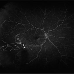 Retinal Hemorrhages
Mar 10 2021 by Kachelle Brown
Ultra widefield Fluorescein Angiography of a 48-year-old female with retinal hemorrhages affecting her right eye. Physician suspect sickle cell due to family history, and has ordered labs to rule out.
Photographer: Kachelle Brown
Imaging device: Optos California
Condition/keywords: Optos, retinal hemorrhage, sickle cell retinopathy, ultra-wide field imaging
-
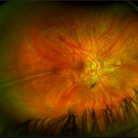 Prominent Long Ciliary Nerve
Jan 25 2022 by Kachelle Brown
Ultra-wide field photograph of a 48-year-old female with a prominent long ciliary nerve. Patient presented asymptomatic, and was referred for a macula on retinal detachment. Patient was diagnosed with high myopia and a posterior vitreous detachment, and the physician discussed increased risk of floaters, myopic degeneration and retinal detachment associated with high myopia. -24.00 prior to cataract surgery OU per patient.
Photographer: Kachelle Brown
Imaging device: Optos California
Condition/keywords: fundus photograph, high myopia, long ciliary nerve, optos, right eye, ultra-widefield image
-
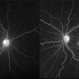 Cystoid Macular Degeneration
Feb 1 2023 by Kachelle Brown
Fluorescein Angiogram of a 56 year old woman with bilateral Cystoid Macular Degeneration. Patient vision was 20/60 OU.
Photographer: Kachelle Brown OMA, Retina Specialist of Michigan
Condition/keywords: cystoid macular degeneration, cystoid macular edema (CME), FA late phase
A project from the American Society of Retina Specialists