-
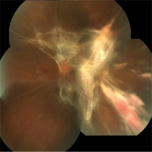 Extensive Tractional Retinal Detachment in Proliferative Diabetic Retinopathy
Jun 4 2018 by Diva Kant Misra, MBBS, DO, DNB, MNAMS, FVRS
Montage fundus photograph of a 54-year-old male diabetic patient showing extensive TRD with PDR.
Photographer: DIVA KANT MISRA
Condition/keywords: diabetes, proliferative diabetic retinopathy (PDR), tractional retinal detachment
-
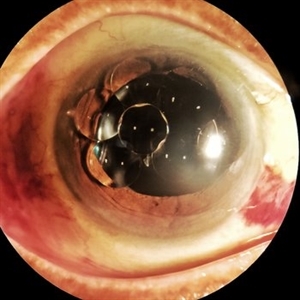 Multiple Silicone Oil Bubbles in Anterior Chamber
Jun 13 2018 by Diva Kant Misra, MBBS, DO, DNB, MNAMS, FVRS
A 56-year-old female patient operated for rhegmatogenous retinal detachment, was found to have multiple silicone oil bubbles in anterior chamber.
Photographer: DIVA KANT MISRA
Condition/keywords: silicone oil
-
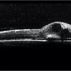 BRVO with Center Involving Cystoid Macular Edema
Nov 5 2018 by Diva Kant Misra, MBBS, DO, DNB, MNAMS, FVRS
OCT image of a 55-year-old man with BRVO and center involving cystoid macular edema, showing a perfect circular cystoid space.
Photographer: HITESHWAR SAIKIA
Condition/keywords: branch retinal vein occlusion (BRVO), clinically significant macular edema (CSME)
-
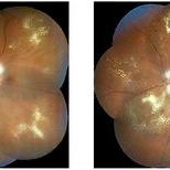 NPDR With Myelinated Nerve Fibers
Nov 5 2018 by Diva Kant Misra, MBBS, DO, DNB, MNAMS, FVRS
Bilateral montage funds photo images of a 56-year-old diabetic patient showing signs of NPDR along with myelinated nerve fibers.
Photographer: Hiteshwar Saikia
Condition/keywords: diabetes, hard exudates, myelinated nerve fibers, nonproliferative diabetic retinopathy
-
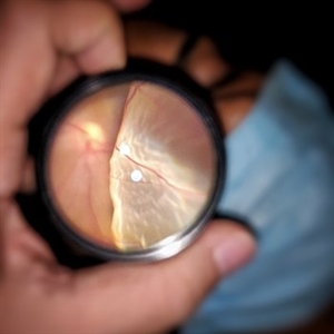 Rhegmatogenous Retinal Detachment
Dec 22 2020 by Diva Kant Misra, MBBS, DO, DNB, MNAMS, FVRS
Fundus photograph of a 35-year-old female with a bullous rhegmatogenous retinal detachment.
Photographer: Diva Kant Misra
Imaging device: Smart Phone ( One Plus 6T)
Condition/keywords: smartphone fundus photography
A project from the American Society of Retina Specialists