-
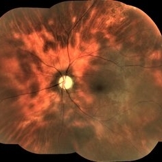
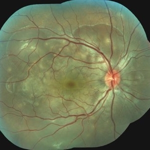 Vogt-Koyanagi-Harada Disease OD
Jun 18 2020 by Renata Garcia Franco, Md
Female 25-year-old with serous retinal detachment in both eyes.
Photographer: Fatima Hernandez
Imaging device: Visucam
Condition/keywords: Vogt-Koyanagi-Harada
-
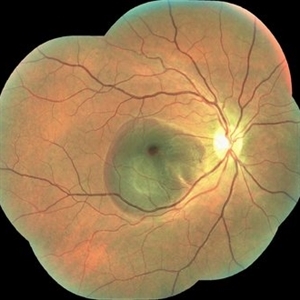
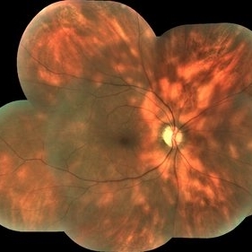
 Vogt-Koyanagi-Harada Disease OS
Jun 18 2020 by Renata Garcia Franco, Md
Female 25-year-old with serous retinal detachment in both eyes.
Photographer: Fatima Hernandez
Imaging device: Visucam
Condition/keywords: Vogt-Koyanagi-Harada
-
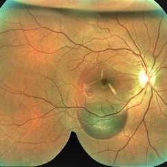
 Traumatic Submacular Hemorrhage
Jul 17 2020 by Renata Garcia Franco, Md
Fundus photograph of 18-year-old male, who suffered trauma to the right eye 7 days before. Choroidal rupture was visible nasal to the fovea.
Photographer: Fatima Hernandez, Instituto de la Retina del Bajio SC
Imaging device: Zeiss
Condition/keywords: blunt, submacular hemorrhage, trauma
-
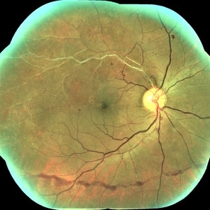
 Pneumatic Displacement of Submacular Haemorrhage
Jul 17 2020 by Renata Garcia Franco, Md
Fundus photograph of 18-year-old male, who suffered trauma to the right eye 7 days before. SF6 was injected with improved vision the next day, from counting fingers to 20/200. Choroidal rupture with macular involvement.
Photographer: Fatima Hernandez, Instituto de la Retina del Bajio SC
Imaging device: Zeiss
Condition/keywords: blunt trauma, submacular hemorrhage
-
 Mixed Occlusion of Artery and Vein
Jan 6 2021 by Renata Garcia Franco, Md
Male with a history of smoking, sudden low vision of the right eye, retinal neovascularization and inferior preretinal hemorrhage.
Photographer: Fatima Hernandez, Instituto de la Retina del Bajio SC
Imaging device: Zeiss
Condition/keywords: arterial occlusion
-
 Central Retinal Artery Occlusion
Jan 22 2021 by Renata Garcia Franco, Md
65-year-old male, history of uncontrolled systemic arterial hypertension. Fluorescein angiography (FA) shows a delay in filling of the retinal arteries.
Photographer: Fatima Hernandez, Instituto de la Retina del Bajio SC
Imaging device: Zeiss
Condition/keywords: central retinal artery occlusion (CRAO)
-
 Central Retinal Artery Occlusion
Jan 22 2021 by Renata Garcia Franco, Md
65-year-old male, history of uncontrolled systemic arterial hypertension. Segmentation of blood in retinal arterioles, retinal whitening and cherry red spot.
Photographer: Fatima Hernandez, Instituto de la Retina del Bajio SC
Imaging device: Zeiss
Condition/keywords: central retinal artery occlusion (CRAO)
-
 Birdshot Retinochoroidopathy
Jan 22 2021 by Renata Garcia Franco, Md
50-year-old female with history of floaters and hazy vision. Yellow-white lesions in the fundus, retinal vasculitis, epiretinal membrane and cystoid macular edema.
Photographer: Fatima Hernandez, Instituto de la Retina del Bajio SC
Imaging device: Zeiss
Condition/keywords: birdshot retinochoroidopathy
-
 Birdshot Retinochoroidopathy
Jan 22 2021 by Renata Garcia Franco, Md
50 -year-old female with history of floaters and hazy vision. Yellow-white lesions in the fundus, retinal vasculitis and 2+ vitreous haze.
Photographer: Fatima Hernandez, Instituto de la Retina del Bajio SC
Imaging device: Zeiss
Condition/keywords: birdshot retinochoroidopathy
-
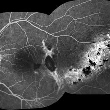 Traumatic Macular Hole And Bruch Membrane Rupture
Jan 22 2021 by Renata Garcia Franco, Md
FA shows hypofluorescence in early frames due to a break in choriocapillaris and choroidal vessels at the rupture site with staning at late phases.
Photographer: Fatima Hernandez, Instituto de la Retina del Bajio SC
Imaging device: Zeiss
Condition/keywords: traumatic macular hole
-
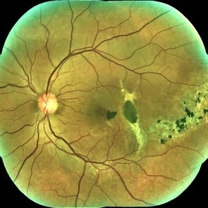 Traumatic Macular Hole And Bruch Membrane Rupture
Jan 22 2021 by Renata Garcia Franco, Md
Male with history of ocular blunt injury, full-thickness macular hole, RPE changes and Bruch membrane rupture.
Photographer: Fatima Hernandez, Instituto de la Retina del Bajio SC
Imaging device: Zeiss
Condition/keywords: traumatic macular hole
-
 Relentless Placoid Chorioretinitis
Jan 22 2021 by Renata Garcia Franco, Md
20-year-old male with reduction of vision in both eyes, scotoma and metamorphopsia. Widespread multiple chorioretinal lesions with RPE hyperplasia, which appear from posterior pole to peripheral retina.
Photographer: Fatima Hernandez, Instituto de la Retina del Bajio SC
Imaging device: Zeiss
Condition/keywords: chorioretinitis
-
 Relentless Placoid Chorioretinitis
Jan 22 2021 by Renata Garcia Franco, Md
20-year-old male with reduction of vision in both eyes, scotoma and metamorphopsia. Widespread multiple chorioretinal lesions with RPE hyperplasia, which appear from posterior pole to peripheral retina and inactive choroidal neovascular membrane.
Photographer: Fatima Hernandez, Instituto de la Retina del Bajio SC
Imaging device: Zeiss
Condition/keywords: chorioretinitis
-
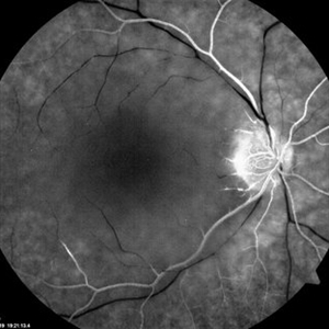 Central Retinal Artery Occlusion
Mar 2 2021 by Renata Garcia Franco, Md
Fundus fluorescein angiography in the acute phase reveals normal choroidal filling with delayed or absent filling of the central retinal artery.
Photographer: Guillermina Hernandez
Imaging device: Zeiss
Condition/keywords: central artery
-
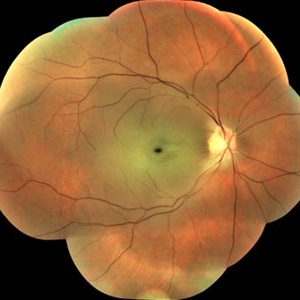 Central Retinal Artery Occlusion
Mar 2 2021 by Renata Garcia Franco, Md
Retinal edema, cherry spot, retinal arteriolar attenuation and segmentation of blood in retinal arterioles.
Photographer: Guillermina Hernandez
Imaging device: Zeiss
Condition/keywords: central artery
A project from the American Society of Retina Specialists