-
 Lupus Hemorrhagic Occlusive Vasculitis
Apr 23 2018 by Frank Chin
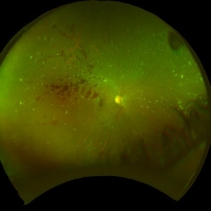
Fundus photograph of the right eye of a 24-year-old woman with history of systemic lupus erythematosus who presented with decreased visual acuity for 2-3 days found to have lupus hemorrhagic occlusive vasculitis with mild disc elevation, diffuse punctate cotton wool spots and dot blot hemorrhages, and a hemorrhage occlusive vasculitis along the superior branch of the superotemporal arcade involving the artery and vein.
Photographer: Frank Chin, MD, George Washington University
Imaging device: Optos 200Tx
Condition/keywords: blot hemorrhages, cotton wool spots, occlusive vasculitis, systemic lupus erythematosus (SLE) vasculitis
A project from the American Society of Retina Specialists