-
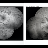
 Valsalva Retinopathy
Apr 5 2018 by Mohamed Tawfik, MD
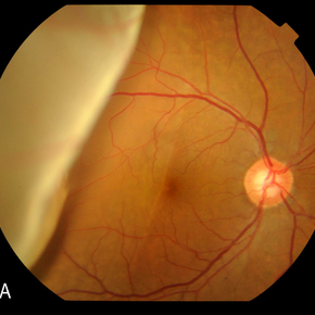
Fundus photography of 30-year-old male presented with sudden drop of vision .. diagnosed as valsalva retinopathy .. we do YAG hylodotomy ... vision is improved from HM to 1.0.
Photographer: Mohamed A.Tawfik MD,FRCSed
Condition/keywords: valsalva retinopathy
-
 Dropped IOL
Apr 5 2018 by Mohamed Tawfik, MD
Intra operative photo of a case of dropped IOL after phaco+IOL.
Photographer: Mohamed A,Tawfik
Imaging device: Intra operative Photography Screen shoot
Condition/keywords: dropped intraocular lens (IOL)
-
 Sub Total Retinal Detachment
Apr 5 2018 by Mohamed Tawfik, MD
Fundus photography of a case of sub total RD macular OFF fovea ON with BCVA 1.0.
Photographer: Mohamed A,TAwfik
-
 Giant Retinal Tear
Apr 5 2018 by Mohamed Tawfik, MD
Intra-operative photos of giant tear repair in pseudophakic eye.
Photographer: Mohamed A.Tawfik
Imaging device: Intra opeative Photography Screen shoot
Condition/keywords: giant retinal tear
-
 Dropped Lens and IOLs
Apr 5 2018 by Mohamed Tawfik, MD
Group photo of dropped crystalline lens and IOLs.
Photographer: Mohamed A.Tawfik MD , FRCSed
Imaging device: intra opeative Photography Screen shoot
Condition/keywords: dropped intraocular lens (IOL), dropped nucleus
-
 Focal Macular Edema
Apr 5 2018 by Mohamed Tawfik, MD
FAF of a case of Focal Macular edema treated with modified grid laser.
Photographer: Mohamed A,Tawfik MD,FRCSed
Condition/keywords: cystoid macular edema (CME)
-
 Dropped Capsular IOL Bag Complex
Apr 5 2018 by Mohamed Tawfik, MD
A case of high myope with dropped capsular IOL bag complex.
Photographer: Mohamed A,Tawfik
Imaging device: intra opeative Photography Screen shoot
Condition/keywords: dropped capsular IOL bag complex
-
 Disc Collateral
Apr 5 2018 by Mohamed Tawfik, MD
A case of Old CRVO presented with disc collateral.
Photographer: Mohamed A,Tawfik MD , FRCSed
Condition/keywords: central retinal vein occlusion (CRVO), disc
-
 ILM Removal
Apr 5 2018 by Mohamed Tawfik, MD
Steps Of ILM peel stained with brilliant blue under PFO.
Photographer: Mohamed A,Tawfik MD,FRSCed
Imaging device: intra opeative Photography Screen shoot
Condition/keywords: internal limiting membrane (ILM) peeling
-
 Sub Total RD
Apr 5 2018 by Mohamed Tawfik, MD
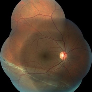
Fundus Photo Of a case of Sub-total RD macular ON BCVA 1.0; demonstrated demarcation line denote old RD.
Photographer: Moahmed A,Tawfik MD , FRCSed
-
 Macular Hole RD
Apr 5 2018 by Mohamed Tawfik, MD
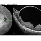
OCT image of a case of macular hole RD show improved of anatomical view with improving of vision.
Photographer: Mohamed A,Tawfik MD , FRCSed
Condition/keywords: macular hole
-
 Silicon Bubble In AC
Apr 5 2018 by Mohamed Tawfik, MD
image of a case post phaco vitrectomy show silicon bubble in anterior chamber.
Photographer: Mohamed A , Tawfik MD , FRCSed
Imaging device: Mobile Photography
Condition/keywords: silicone oil
-
 Retinal Detachment
Apr 5 2018 by Mohamed Tawfik, MD
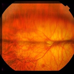
Fundus Photography of 6 day post operative vitrectomy of a case of total RD ON SF6 show fluid level with attached retina.
Photographer: Mohamed A,Tawfik MD,FRCSed
-
 Pan-Retinal Photocoagulation
Apr 5 2018 by Mohamed Tawfik, MD
Wide field FFA post phaco vitrectomy of a case of vitreous hemorrhage show PRP with regression of diabetic retinopathy.
Photographer: Mohamed A,Tawfik MD,FRCSed
Condition/keywords: pan-retinal photocoagulation (PRP)
-
 Fibrovascular Membrane
Apr 5 2018 by Mohamed Tawfik, MD
16 mm wide field OCT scan of a case of fiber-vascular membrane demonstrate the point of attachment of membrane.
Photographer: Mohamed A,Tawfik MD,FRCSed
Condition/keywords: fibrotic neovascularization, fibrous proliferation, fibrovascular change
-
 Proliferative Diabetic Retinopathy
Apr 5 2018 by Mohamed Tawfik, MD
Fundus autofluorescence of a case of proliferative diabetic retinopathy demonstrate the new vessels on disc and elsewhere.
Photographer: Mohamed A,Tawfik MD,FRCSed
Condition/keywords: proliferative diabetic retinopathy (PDR)
-
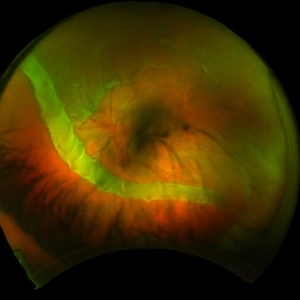 Giant Retinal Tear
Feb 19 2022 by Mohamed Tawfik, MD
wide filed image show a case of 180 degree giant retinal detachment
Photographer: Mohamed tawfik
Condition/keywords: detachment
A project from the American Society of Retina Specialists