-
 Fundus Photo IRVAN
Feb 7 2021 by Natalie Huang, MD
Color fundus photography of a 31-year-old man with idiopathic retinitis, vasculitis, aneurysms and neuro-retinitis (IRVAN) syndrome.
Photographer: Judy, Kaplan, SUNY Upstate Medical University, Department of Ophthalmology and Visual Science
Imaging device: Zeiss Clarus 500
Condition/keywords: IRVAN Syndrome
-
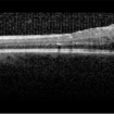 OCT-IRVAN
Feb 7 2021 by Natalie Huang, MD
Optic coherence tomography of a 31-year-old man with idiopathic retinitis, vasculitis, aneurysms and neuro-retinitis (IRVAN) syndrome.
Photographer: Judy, Kaplan, SUNY Upstate Medical University, Department of Ophthalmology and Visual Science
Imaging device: Heidelberg Spectralis
Condition/keywords: IRVAN Syndrome
-
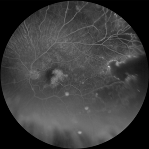 FA1-IRVAN
Feb 7 2021 by Natalie Huang, MD
Fluorescence angiography of a 31-year-old man with idiopathic retinitis, vasculitis, aneurysms and neuro-retinitis (IRVAN) syndrome.
Photographer: Judy, Kaplan, SUNY Upstate Medical University, Department of Ophthalmology and Visual Science
Imaging device: Heidelberg Spectralis
Condition/keywords: IRVAN Syndrome
-
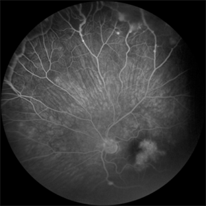 FA2-IRVAN
Feb 7 2021 by Natalie Huang, MD
Fluorescence angiography of a 31-year-old man with idiopathic retinitis, vasculitis, aneurysms and neuro-retinitis (IRVAN) syndrome.
Photographer: Judy, Kaplan, SUNY Upstate Medical University, Department of Ophthalmology and Visual Science
Imaging device: Heidelberg Spectralis
Condition/keywords: IRVAN Syndrome
A project from the American Society of Retina Specialists