-
 IOL
IOL
Jan 17 2018 by Emily Cooper
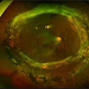
Optos image of 47-year-old man with a now worsening retinal detachment that had been treated by pneumatic retinopexy.
Photographer: Emily Cooper, Retina Specialists of Michigan
Imaging device: Optos Ultra Wide Field
Condition/keywords: chronic retinal detachment, intraocular lens (IOL)
-
 Retinitis Pigmentosa
Jan 17 2018 by Emily Cooper
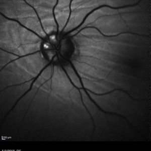
Fundus Photos of 11-year-old with retinitis pigmentosa.
Photographer: Emily Cooper, Retina Specialists of Michigan
Imaging device: Optos Ultra Wide Field
Condition/keywords: retinectomy, retinitis pigmentosa
-
 Choroidal Folds and Optic Disc Drusen
Aug 1 2018 by Emily Cooper
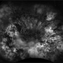
Fundus autofluorescence photo of a 62-year-old man who presented for evaluation of choroidal folds and optic disc drusen. He is currently following up with neuro-ophthalmology and has suspected intracranial hypertension.
Photographer: Emily Cooper, Retina Specialists of Michigan
Condition/keywords: choroidal folds, drusen of optic disc
-
 Choroidal Detachment
Oct 4 2018 by Emily Cooper
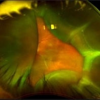
Optos photograph of an 80-year-old man presenting with red, painful eye after heart surgery.
Photographer: Emily Cooper, Retina Specialists of Michigan, Grand Rapids MI
Imaging device: Optos
Condition/keywords: choroidal detachment, posterior scleritis
-
 Coats' Disease
May 21 2019 by Emily Cooper
Fundus photograph of an 73-year-old man with Coats' Disease.
Photographer: Emily Cooper, Retina Specialists of Michigan
Imaging device: Optos
Condition/keywords: Coats' disease
A project from the American Society of Retina Specialists