-
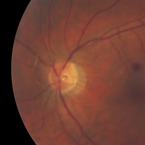
 Elmiron Toxicity
Jan 12 2018 by Jessica Norkus
Bilateral ultra-wide field pseudo-color and autofluorescent images of a 46-year-old female with Elmiron Toxicity.
Photographer: Jessica Norkus
Imaging device: Optos
Condition/keywords: autofluorescence imaging, bilateral, color fundus photograph, drug toxicity, Optos, toxic maculopathy, toxic retinopathy, ultra-wide field imaging
-
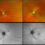
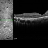 Pigmentary Retinal Dystrophy
Mar 29 2019 by Jessica Norkus
Heidelberg Spectralis image of 41-year-old male patient with pigmentary retinal dystrophy. Atypical findings due to unilateral presentation. Patient has been experiencing symptoms for 15 years, notes significant nyctalopia.
Photographer: Jessica Norkus
Imaging device: Heidelberg Spectralis
Condition/keywords: bone spicule, Heidelburg Spectralis, optical coherence tomography (OCT), pigment changes, unilateral blindness
-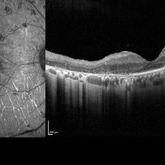
 Pigmentary Retinal Dystrophy
Mar 29 2019 by Jessica Norkus
Optos ultra wide field image of 41-year-old male patient with pigmentary retinal dystrophy. Atypical findings due to unilateral presentation. Patient has been experiencing symptoms for 15 years, notes significant nyctalopia.
Photographer: Jessica Norkus
Imaging device: Optos Ultra Wide Field Camera
Condition/keywords: abnormal fundus, bone spicule, color fundus photograph, color photo, fundus autofluorescence (FAF), fundus photograph, Optos, peripheral bone spicules, pigment changes, ultra-wide field imaging, unilateral blindness
-
 Pigmentary Retinal Dystrophy
Mar 29 2019 by Jessica Norkus
Optos ultra wide field image of 41-year-old male patient with pigmentary retinal dystrophy. Atypical findings due to unilateral presentation. Patient has been experiencing symptoms for 15 years, notes significant nyctalopia.
Photographer: Jessica Norkus
Imaging device: Optos Ultra Wide Field Camera
Condition/keywords: abnormal fundus, bone spicule, color fundus photograph, color photo, fundus photograph, Optos, peripheral bone spicules, pigment changes, ultra-wide field imaging, unilateral blindness
-
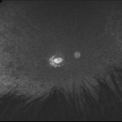
 Weiss Ring
Oct 22 2019 by Jessica Norkus
Fundus photo taken on TopCon TRC 50Dx camera of a 60-year-old patient who has experienced an acute PVD. Chief complaint of "large floater" OS prompted exam. Physician noted a significant Weiss Ring and requested fundus color photo for documentation.
Photographer: Jessica Norkus, COA (Retina Specialists of Michigan)
Imaging device: TopCon TRC 50Dx
Condition/keywords: color fundus photograph, color photo, fundus photograph, nerve, optic disc, posterior vitreous detachment, retina, vitreous floaters, Weiss ring
-
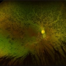
 Congenital Hypertrophy of the Retinal Pigment Epithelium
Nov 11 2019 by Jessica Norkus
Bilateral Optos ultra wide field imaging of a 31-year-old female patient with CHRPE lesions. Lesions in OD were suspicious of Gardner Syndrome due to familial history of cancerous polyps in colon. Patient underwent colonoscopy and was deemed clear.
Photographer: Jessica Norkus, COA, Retina Specialists of Michigan
Imaging device: Optos Ultra Wide Field Camera
Condition/keywords: bear tracks, bilateral, color fundus photograph, color photo, congenital hypertrophy of the retinal pigment epithelium (CHRPE), fundus autofluorescence (FAF), fundus photograph, lacunae, macula, optic disc, Optos, pseudocolor, ultra-wide field imaging
A project from the American Society of Retina Specialists