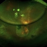
-
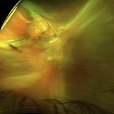 Pre-OP
Jan 7 2018 by John S. King, MD
61-year-old developed a large choroidal OD after a bleb revision. 30 years ago had an IOFB removed. The initial photos, POD 1 and POW 3 photos are provided. Dr. Zocchi performed the surgery. Surgery included 42 band placed 360 (superior bleb was fibrosed and left in place during conj peritomy); suprachoroidal drainiage was via radial incision; a pre-existing ST tear was present; the retinal adhesions were removed with blunt dissection, then remaining pvr was peeled; PFO used to flatten retina; 5000 cs sil oil used. Post-op vision has improved for near to J5.
Imaging device: Optos
Condition/keywords: suprachoroidal hemorrhage
-
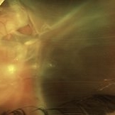 Pre-OP
Jan 7 2018 by John S. King, MD
61-year-old developed a large choroidal OD after a bleb revision. 30 years ago had an IOFB removed. The initial photos, POD 1 and POW 3 photos are provided. Dr. Zocchi performed the surgery. Surgery included 42 band placed 360 (superior bleb was fibrosed and left in place during conj peritomy); suprachoroidal drainiage was via radial incision; a pre-existing ST tear was present; the retinal adhesions were removed with blunt dissection, then remaining pvr was peeled; PFO used to flatten retina; 5000 cs sil oil used. Post-op vision has improved for near to J5.
Imaging device: Optos
Condition/keywords: suprachoroidal hemorrhage
-
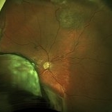 POD 1
POD 1
Jan 7 2018 by John S. King, MD
61-year-old developed a large choroidal OD after a bleb revision. 30 years ago had an IOFB removed. The initial photos, POD 1 and POW 3 photos are provided. Dr. Zocchi performed the surgery. Surgery included 42 band placed 360 (superior bleb was fibrosed and left in place during conj peritomy); suprachoroidal drainiage was via radial incision; a pre-existing ST tear was present; the retinal adhesions were removed with blunt dissection, then remaining pvr was peeled; PFO used to flatten retina; 5000 cs sil oil used. Post-op vision has improved for near to J5.
Imaging device: Optos
Condition/keywords: suprachoroidal hemorrhage
-
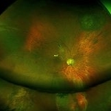 POW 3
POW 3
Jan 7 2018 by John S. King, MD
61-year-old developed a large choroidal OD after a bleb revision. 30 years ago had an IOFB removed. The initial photos, POD 1 and POW 3 photos are provided. Dr. Zocchi performed the surgery. Surgery included 42 band placed 360 (superior bleb was fibrosed and left in place during conj peritomy); suprachoroidal drainiage was via radial incision; a pre-existing ST tear was present; the retinal adhesions were removed with blunt dissection, then remaining pvr was peeled; PFO used to flatten retina; 5000 cs sil oil used. Post-op vision has improved for near to J5.
Imaging device: Optos
Condition/keywords: suprachoroidal hemorrhage
A project from the American Society of Retina Specialists