-
 Roth Spots - Bacterial Endocarditis
Dec 3 2017 by John S. King, MD
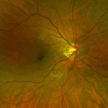
Initial presentation; 29-year-old white female denied ivdu p/c with acute scotoma due to the sub-ILM foveal heme. She did have some roth spots in both eyes. There was a focal area of periphlebitis just superior to the fovea OD. Work up for roth spots and retinal vasculitis initiated. She did have a low grade fever that she attributed to a urinary tract infection being treated by her PCP.
Imaging device: Optos
Condition/keywords: sub-inner limiting membrane hemorrhage, white centered retinal hemorrhage (Roth Spot)
-
 Sub-ILM Heme in patient with Roth Spots - Bacterial Endocarditis
Dec 3 2017 by John S. King, MD
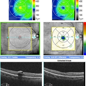
29 yo wf denied ivdu p/c with acute scotoma. OCT shows sub-ILM heme in foveal region (left) 20/300 that resolved spontaneously a few weeks later, back to baseline acuity (right).
Imaging device: Cirrus
Condition/keywords: bacterial endocarditis, sub-inner limiting membrane hemorrhage, white centered retinal hemorrhage (Roth Spot)
-
 Roth Spots - Bacterial Endocarditis
Dec 3 2017 by John S. King, MD
Few weeks later; some areas resolved, and new roth spots; sub-ILM foveal heme in right eye resolved and vision back to baseline; full work-up pending.
Imaging device: Optos
Condition/keywords: bacterial endocarditis, white centered retinal hemorrhage (Roth Spot)
-
 Roth Spots - Bacterial Endocarditis
Dec 3 2017 by John S. King, MD
Two months later; interim was hospitalized for bacterial infection of mitral valve; roth spots resolved.
Imaging device: Optos
Condition/keywords: bacterial endocarditis, white centered retinal hemorrhage (Roth Spot)
A project from the American Society of Retina Specialists