-
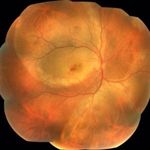 Acute Syphilitic Posterior Placoid Chorioretinitis
Sep 3 2016 by ADRIANO FERREIRA
66-year-old woman with acute visual acuity loss.
Photographer: Claudio Zett Lobo, UNIFESP
Condition/keywords: acute syphilitic posterior placoid chorioretinitis
-
 Acute Syphilitic Posterior Placoid Chorioretinitis
Sep 3 2016 by ADRIANO FERREIRA
66 - year -old woman with acute visual acuity loss.
Photographer: Claudio Zett Lobo, UNIFESP
Condition/keywords: acute syphilitic posterior placoid chorioretinitis
-
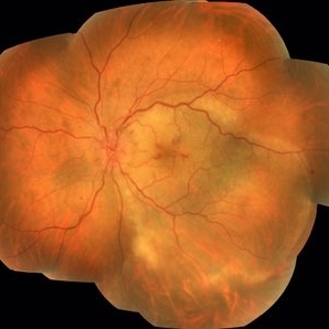
 Acute Syphilitic Posterior Placoid Chorioretinitis
Sep 3 2016 by ADRIANO FERREIRA
66-year-old woman with acute visual acuity loss.
Photographer: Claudio Zett Lobo, UNIFESP
Condition/keywords: acute syphilitic posterior placoid chorioretinitis
-
 Acute Syphilitic Posterior Placoid Chorioretinitis
Sep 3 2016 by ADRIANO FERREIRA
66-year-old woman with acute visual acuity loss.
Photographer: Claudio Zett Lobo, UNIFESP
Condition/keywords: acute syphilitic posterior placoid chorioretinitis
-
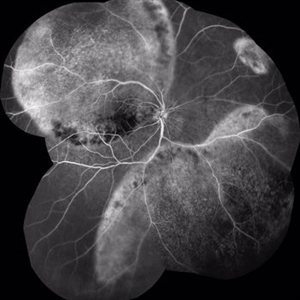 Acute Syphilitic Posterior Placoid Chorioretinitis
Sep 3 2016 by ADRIANO FERREIRA
66-year-old woman with acute visual acuity loss.
Photographer: Claudio Zett Lobo, UNIFESP
Imaging device: Intravenous Fluorescein angiography
Condition/keywords: acute syphilitic posterior placoid chorioretinitis
-
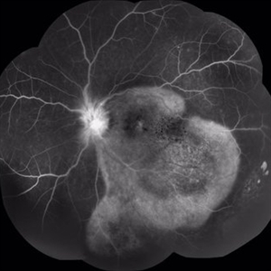 Acute Syphilitic Posterior Placoid Chorioretinitis
Sep 3 2016 by ADRIANO FERREIRA
66-year-old woman with acute visual acuity loss.
Photographer: Claudio Zett Lobo
Imaging device: Intravenous Fluorescein angiography
Condition/keywords: acute syphilitic posterior placoid chorioretinitis
-
 Acute Necrotizing Retinal Vasculitis as Onset of Systemic Lupus Erythematosus.
Sep 3 2016 by ADRIANO FERREIRA
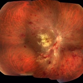
A 28-year-old white man was referred to the rheumatology clinic with gradually and rapid deterioration of the vision (both eyes). In this picture, we can observe cotton wool spots in the papillomacular area and extensive hemorrhages in posterior polo and in the middle periphery. Hard exudates are present in macular area (macular edema)
Photographer: Claudio Zett Lobo
Imaging device: TRC50DXi TOPCON
Condition/keywords: systemic lupus erythematosus (SLE) vasculitis, vasculitis
-
 Acute Necrotizing Retinal Vasculitis as Onset of Systemic Lupus Erythematosus.
Sep 3 2016 by ADRIANO FERREIRA
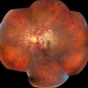
A 28-year-old white man was referred to the rheumatology clinic with gradually and rapid deterioration of the vision (both eyes). In this picture we can observe cotton wool spots in the papillomacular area and extensive hemorrhages in the left eye.
Photographer: Claudio Zett Lobo
Imaging device: TRC50DXi TOPCON
Condition/keywords: systemic lupus erythematosus (SLE) vasculitis, vasculitis
-
 Acute Necrotizing Retinal Vasculitis as Onset of Systemic Lupus Erythematosus.
Sep 3 2016 by ADRIANO FERREIRA
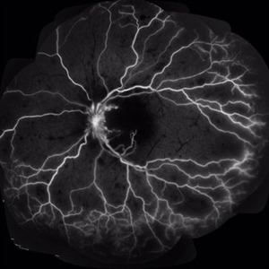
28-year-old white man was referred to the rheumatology clinic with gradually and rapid deterioration of the vision (both eyes). In this picture, we can observe vasculitis (leakage from vessels) and diffuse ischemia in the left eye.
Photographer: Claudio Zett Lobo
Imaging device: HRA-Spectralis
Condition/keywords: systemic lupus erythematosus (SLE) vasculitis, vasculitis
-
 Ophthalmic Artery Occlusion
Jan 28 2017 by ADRIANO FERREIRA
32-year-old female with a sudden loss of vision in the left eye. This fundus photograph shows central arterial occlusion plus central vein occlusion secondary to ophthalmic artery occlusion. This patient has thromboangitis obliterans.
Photographer: Mr. Jose Luiz
Imaging device: TRC50DXi
Condition/keywords: arterial occlusion, occlusion of retinal vein
-
 Retinal Angiomatous Proliferation
Jan 29 2017 by ADRIANO FERREIRA
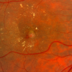
Fundus photograph of an 56-year-old man with a retinal angiomatous proliferation (RAP). RAP has been known as a variant of exudative age-related macular degeneration (AMD) with a unfavorable prognosis.
Photographer: Laercio
Condition/keywords: vascular anomaly
-
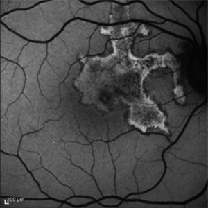 Serpiginous Choroiditis
May 13 2017 by ADRIANO FERREIRA
Autofluorescence imaging of an 48-year-old man with decreased visual acuity in the right eye for 15 days. In time, undergoing tuberculosis treatment.
Photographer: Claudio Zetts Lobo
Condition/keywords: autofluorescence imaging, serpiginous choroiditis, tuberculosis
-
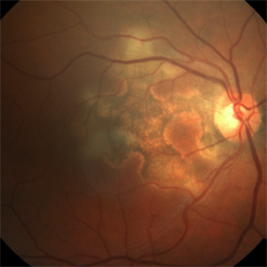 Serpiginous Choroiditis
May 13 2017 by ADRIANO FERREIRA
Fundus photograph of an 48-year-old man with decreased visual acuity in the right eye for 15 days. In time, undergoing tuberculosis treatment.
Photographer: Claudio Zetts Lobo
Imaging device: TRC50DXi
Condition/keywords: serpiginous choroiditis, tuberculosis
-
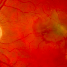 Choroidal Rupture and Secondary Choroidal Neovascularization
May 13 2017 by ADRIANO FERREIRA
Fundus photograph of an 32-year-old man with visual acuity decrease in left eye 30 days ago. Previous ophthalmologic history of blunt ocular trauma in this eye 2 years ago. At funduscopic examination we observe choroidal rupture with subretinal neovascular membrane.
Photographer: Jose Luiz
Condition/keywords: blunt trauma, choroidal neovascularization (CNV), choroidal rupture
-
 Macular Hole and Retinoschisis in Goldmann - Favre Syndrome
May 13 2017 by ADRIANO FERREIRA
Fundus photograph of an 9-year-old child with Goldmann-Favre syndrome presenting with a macular hole and retinoschisis in the right eye.
Photographer: Jose Luiz
Condition/keywords: full thickness macular hole, Goldmann-Favre Syndrome, retinoschisis
A project from the American Society of Retina Specialists