-
 Retinoblastoma Group-A (International Classification)
Oct 2 2015 by Aparna Ramasubramanian
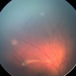
Retcam fundus photography of a child with bilateral sporadic retinoblastoma showing multiple small retinal tumors (superotemporally and superiorly). By the international classification this would be graded as a Group A retinoblasoma (Rb size < 3 mm).
Photographer: Aparna Ramasubramanian
Condition/keywords: retinoblastoma
-
 Retinoblastoma Group-B (International Classification)
Oct 2 2015 by Aparna Ramasubramanian
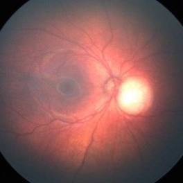
Retcam fundus photography of a child with bilateral sporadic retinoblastoma showing an elevated tumor adjacent to the optic nerve. By the international classification this would be graded as a Group B retinoblasoma (Rb size >3 mm or =3 mm to foveola or =1.5 mm to disc or srf =3 mm from margin).
Photographer: Aparna Ramasubramanian
Condition/keywords: retinoblastoma
-
 Retinoblastoma Group-C (International Classification)
Oct 2 2015 by Aparna Ramasubramanian
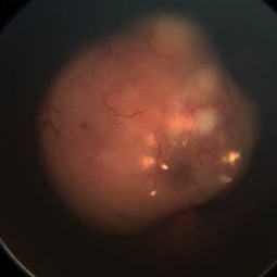
Retcam fundus photography of a child with unilateral sporadic retinoblastoma showing an elevated tumor overhanging the optic nerve and macula. By the international classification this would be graded as a Group C retinoblasoma (subretinal or vitreous seeds < 3 mm from the margin of the tumor).
Photographer: Aparna Ramasubramanian
Condition/keywords: retinoblastoma
-
 Retinoblastoma Group-D (International Classification)
Oct 2 2015 by Aparna Ramasubramanian
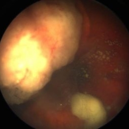
Retcam fundus photography of a child with unilateral sporadic retinoblastoma showing the elevated nasal tumor with diffuse vitreous seeds. By the international classification this would be graded as a Group D retinoblasoma (subretinal or vitreous seeds > 3 mm from the margin of the tumor).
Photographer: Aparna Ramasubramanian
Condition/keywords: retinoblastoma
-
 Retinoblastoma Group-E (International Classification)
Oct 2 2015 by Aparna Ramasubramanian
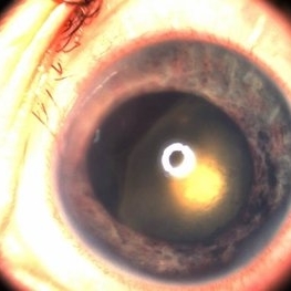
Retcam fundus photography of a child with unilateral sporadic retinoblastoma showing the tumor close to the lens and iris neovascularization with high intraocular pressure. By the international classification this would be graded as a Group E retinoblasoma.
Photographer: Aparna Ramasubramanian
Condition/keywords: retinoblastoma
-
 Retinoblastoma Ultrasound
Oct 2 2015 by Aparna Ramasubramanian
Ultrasonography of a retinoblastoma tumor shows hyperreflective echoes suggestive of calcification. It is seen in 90% of retinoblastoma patients and is an important diagnostic sign.
Photographer: Aparna Ramasubramanian
Condition/keywords: A-scan ultrasound, B scan ultrasound, calcification, retinoblastoma
A project from the American Society of Retina Specialists