-
 Optic Nerve Pit and Retinal Detachment
Aug 24 2015 by Young Hee Yoon, MD, PhD
Wide fundus photograph of a 44-year-old man. There was optic nerve pit and associated foveal detachment with multiple pigmentation. His best-corrected visual acuity was count finger in 30cm.
Photographer: Yoon Goo Lee, Asan Medical center
Imaging device: Optos
Condition/keywords: optic nerve pit
-
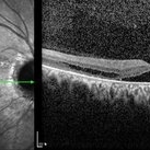 OCT Image of Optic Nerve Pit and Retinal Detachment
Aug 24 2015 by Young Hee Yoon, MD, PhD
OCT image of a 44-year-old man. There was optic nerve pit and associated foveal detachment. His best-corrected visual acuity was count finger in 30cm.
Photographer: Jung Im Cho, Asan Medical Center
Imaging device: Spectralis OCT
Condition/keywords: optic nerve pit
-
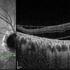
 OCT Image of Optic Nerve Pit and Retinal Detachment
Aug 24 2015 by Young Hee Yoon, MD, PhD
SD-OCT image of a 44-year-old man. There was optic nerve pit and associated foveal detachment. His best-corrected visual acuity was count finger in 30cm.
Photographer: Jung Im Cho, Asan Medical Center
Imaging device: Spectralis OCT
Condition/keywords: optic nerve pit
-
 OCT Image of Optic Nerve Pit and Retinal Detachment
Aug 24 2015 by Young Hee Yoon, MD, PhD
OCT image of a 44-year-old man. There was optic nerve pit and associated foveal detachment. His best-corrected visual acuity was count finger in 30cm.
Photographer: Jung Im Cho, Asan Medical Center
Imaging device: Spectralis OCT
Condition/keywords: optic nerve pit
A project from the American Society of Retina Specialists