-
 Neurocysticercosis
Sep 10 2020 by Anamika Dwivedi
MRI images of a 22-year-old male with h/o headache for 1 month. MRI brain axial scan showing multiple tiny cystic lesions scattered all over the cerebral parenchyma. Post gadolinium contrast coronal scan showing multiple cystic lesion scattered in cerebral parenchyma, few of the lesion showing ring enhancement.
Condition/keywords: neurocysticercosis
-
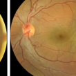 Subretinal Cysticercosis Post Vitrectomy
Sep 10 2020 by Anamika Dwivedi
Fundus photograph of a 22-year-old male, case of subretinal cysticercosis, after PPV and cyst removal showing laser scar at the site of the previous cyst.
Photographer: Dr Anamika Dwivedi
Imaging device: topcon
Condition/keywords: bilateral subretinal cysticercosis
A project from the American Society of Retina Specialists