-
 Lignocaine Retinal Toxicity
Aug 18 2015 by Mallika Goyal, MD
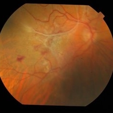
Right eye fundus of a 70-year-old male 3 weeks after inadvertent globe penetration with peribulbar anaesthesia needle and intraocular injection of lignocaine. There is a thick taut epimacular membrane with severely increased central retinal thickness. Fluorescein angiography revealed an occluded retinal arteriole at the macula indicating macular ischaemia underlying the membrane.
Photographer: Mallika Goyal, MD, Apollo Health City, Jubilee Hills, Hyderabad
Condition/keywords: lignocaine retinal toxicity
-
 Lignocaine Retinal Toxicity
Aug 18 2015 by Mallika Goyal, MD
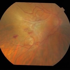
Right eye fundus of a 70-year-old male 3 weeks after inadvertent globe penetration with peribulbar anaesthesia needle and intraocular injection of lignocaine. There is a thick taut epimacular membrane with severely increased central retinal thickness. Fluorescein angiography revealed an occluded retinal arteriole at the macula indicating macular ischaemia underlying the membrane.
Photographer: Mallika Goyal, MD, Apollo Health City, Jubilee Hills, Hyderabad
Condition/keywords: lignocaine retinal toxicity
-
 Lignocaine Retinal Toxicity
Aug 18 2015 by Mallika Goyal, MD
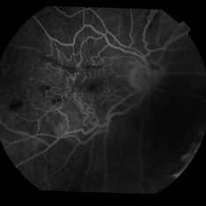
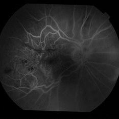
Right eye fluorescein angiogram of a 70-year-old male 3 weeks after inadvertent globe penetration with peribulbar anaesthesia needle and intraocular injection of lignocaine. There is an occluded retinal arteriole indicating macular ischaemia underlying the clinically obvious epimacular membrane.
Photographer: Mallika Goyal, MD, Apollo Health City, Jubilee Hills, Hyderabad
Condition/keywords: lignocaine retinal toxicity
-
 Lignocaine Retinal Toxicity
Aug 18 2015 by Mallika Goyal, MD
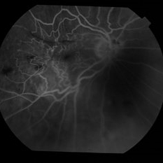
Right eye fluorescein angiogram of a 70-year-old male 3 weeks after inadvertent globe penetration with peribulbar anaesthesia needle and intraocular injection of lignocaine. There is an occluded retinal arteriole indicating macular ischaemia underlying the clinically obvious epimacular membrane.
Photographer: Mallika Goyal, MD, Apollo Health City, Jubilee Hills, Hyderabad
Condition/keywords: lignocaine retinal toxicity
-
 Lignocaine Retinal Toxicity
Aug 21 2015 by Mallika Goyal, MD
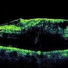
Right eye OCT of a 70-year-old male 3 weeks after inadvertent globe penetration with peribulbar anaesthesia needle and intraocular injection of lignocaine. There is a thick taut epimacular membrane with severely increased central retinal thickness. Fluorescein angiography revealed an occluded retinal arteriole at the macula indicating macular ischaemia underlying the membrane.
Photographer: Mallika Goyal, MD, Apollo Health City, Jubilee Hills, Hyderabad, India
Condition/keywords: lignocaine retinal toxicity
-
 Lignocaine Retinal Toxicity
Aug 21 2015 by Mallika Goyal, MD
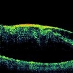
Right eye OCT of a 70-year-old male 3 weeks after inadvertent globe penetration with peribulbar anaesthesia needle and intraocular injection of lignocaine shows a thick taut epimacular membrane with severely increased central retinal thickness. Fluorescein angiography revealed an occluded retinal arteriole at the macula indicating macular ischaemia underlying the membrane.
Photographer: Mallika Goyal, MD, Apollo Health City, Jubilee Hills, Hyderabad, India
Condition/keywords: lignocaine retinal toxicity
-
 Lignocaine Retinal Toxicity
Aug 21 2015 by Mallika Goyal, MD
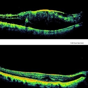
Right eye OCT of a 70-year-old male 3 weeks after inadvertent globe penetration with peribulbar anaesthesia needle and intraocular injection of lignocaine showing a taut epimacular membrane with macular elevation compared to a relatively normal foveal contour immediately after surgery suggesting progressive traction secondary to lignaocaine toxicity. Fluorescein angiography revealed an occluded retinal arteriole at the macula indicating macular ischaemia underlying the membrane.
Photographer: Mallika Goyal, MD, Apollo Health City, Jubilee Hills, Hyderabad, India
Condition/keywords: lignocaine retinal toxicity
A project from the American Society of Retina Specialists